Hercospora tiliae (Pers.) Tul. & C. Tul., Select. fung. carpol. (Paris) 2: 154 (1863), Index Fungorum number: IF 202660
Possible synonyms (See Index Fungorum 2016)
Saprobic on branches and twigs of Tilia sp. Sexual morph: Stromata 700–800 μm wide, prosenchymatous around perithecia, delimited externally by greenish-blackened dense pseudoparenchymatous zone, interior whitish, composed of interwoven hyphae mixed with substrate cells, 3–5 ascomata in a stromata. Ascomata 1–1.05 mm high, 0.24–0.34 mm diam., (x̅ = 1.02 × 334 μm, n = 10), perithecial, small, aggregated, scattered, globose to subglobose, light brown to dark brown, coriaceous, ostiolate, papillate. Papilla 625–645 μm high, 190–290 μm diam., (x̅ = 640 × 250 μm, n = 10), converging and erumpent through stroma surface as single, large opening, wide at the top, narrowing towards the base, dark brown region around base of papilla. Peridium 10–20 μm wide (x̅ = 16 μm, n = 10), comprises light brown, compressed, cells of textura angularis. Asci 140–175 μm × 17–24 μm diam., (x̅ = 160 × 21 μm, n = 10), 8- spored, unitunicate, cylindrical, short-stalked, J- apical apparatus. Ascospores 20–25 μm × 9–11 μm diam., (x̅ = 23 ×10 μm, n = 10), uniseriate, broadly ellipsoid, 1-septate, not or lightly constricted at the septa, hyaline, smooth. Asexual morph: Stromata prosenchymatous. Conidiomata pycnidial, uniloculate, ostiolate, ostiole surrounded by a superficial cap of sterile tissues. Conidiophores elongate. Conidia 14–16.5 × 4.5–6.5 μm (x̅ = 15 × 5 μm, n = 10), hyaline ovoid to ellipsoid, one-celled.
Material examined: SWEDEN. Uppland: Upl. Stockholm: Roslagstull Stockholm, on bark of Tilia sp., L. Romell, 1 April 1887, F148711 (S)
Notes: Hercospora Fr. comprises 15 species (Index Fungorum 2016) with Hercospora tiliae as the type. The characters of the genus include eustromatic, immersed, subepidermal, dark blackish brown, separate, uniloculate, multiloculate or convoluted and thick-walled conidiomata; conidiophores branched extensively at the base, less so above, hyaline, septate, smooth, often developing in mucilage, formed at the base and sides of the conidiomatal wall; and ellipsoid, thick-walled, hyaline, aseptate conidia (18–20 × 6–7.5 µm) (Petrak 1938, Sutton 1980). Phylogenetic studies (Castlebury et al. 2002, Rossman et al. 2007, Voglmayr et al. 2012, Voglmayr & Jaklitsch 2014) based on LSU sequence data, placed H. tiliae in Diaporthales, genera incertae sedis, where it grouped with Melanconis desmazierii. In the present study based on maximum likelihood, maximum parsimony and Bayesian analyses of combined ITS and LSU sequence data, Lamproconium desmazierii (= Melanconis desmazieri) and Hercospora tiliae, clustered in Lamproconiaceae fam. nov.
Fig. 5. Hercospora tiliae (F148711, reference specimen). A Packet of herbarium specimen. B Herbarium specimens. C Cross section of ascomata. D Peridium. E Papilla. F–H Asci in water. I–K Ascospores. Scale bars: C = 200 µm, D, F–H = 40 µm, E = 100 µm.

Fig. 6. Hercospora tiliae (redrawn from Sutton 1980) A Conidia. B Conidiophores and developing conidia. C Conidioma. Scale bars: A = 10 µm, B = 20 µm.
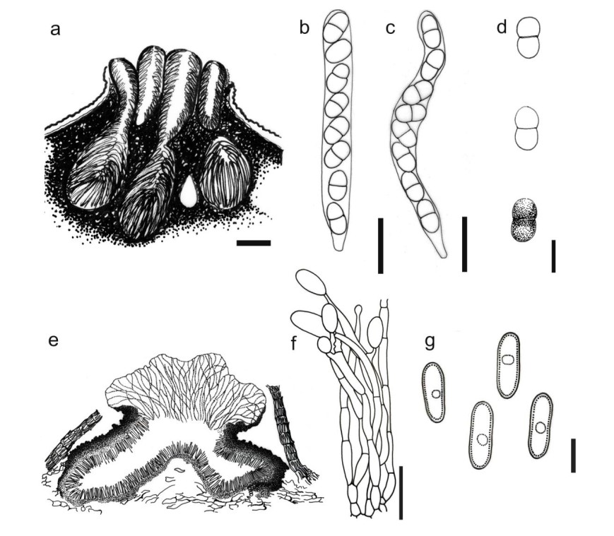
Figure 1 – Hercospora tiliae (redrawn from Sutton 1980 and Norphanphoun et al. 2016). a Ascomata. b, c Asci. d Ascospores. e Conidioma. f Conidiophores and developing conidia. g Conidia. Scale bars: a = 200 μm, b, c = 40 μm, d, f = 20 μm, e = 100 μm, g = 10 μm.

Figure 141 – Hercospora tiliae (redrawn from Sutton 1980 and Norphanphoun et al. 2016). a Ascomata. b, c Asci. d Ascospores. e Conidioma. f Conidiophores and developing conidia. g Conidia. Scale bars: a = 200 μm, b, c = 40 μm, d, f = 20 μm, e = 100 μm, g = 10 μm.