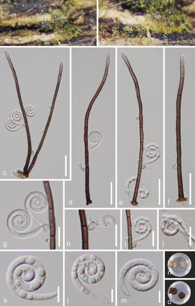
Helicosporium hainanense Y.Z. Lu & J.C. Kang, sp. nov. Fig. 2
MycoBank number: MB; Index Fungorum number: IF; Facesoffungi number: FoF 13101;
Holotype: GZAAS 22-2006
Etymology: ‘hainanense’ referring to collecting site.
Saprobic on decaying woody substrate. Sexual morph Undetermined. Asexual morph Hyphomycetous, helicosporous. Colonies on the substratum superficial, effuse, gregarious, yellow green. Mycelium partly immersed, pale brown to brown, septate, branched hyphae, with masses of crowded, glistening conidia. Conidiophores 118–182 µm long, 2.5–4 µm wide ( = 155 × 3 µm, n = 30), macronematous, mononematous, cylindrical, unbranched, straight or slightly flexuous, septate, pale brown to dark brown, smooth-walled. Conidiogenous cells holoblastic, mono- to polyblastic, discrete, determinate, arising laterally from lower portion of the conidiophores as tiny bladder-like protrusions, 2–8.5 µm long, 1.5–3.5 µm diam., with each bearing 1–3 tiny conidiogenous loci, hyaline to pale brown, smooth-walled. Conidia 11–13 µm diam. and conidial filament 2–3 µm wide ( = 12 × 2.5 µm, n = 20), 55–60 µm long, solitary, pleurogenous, helicoid, tightly coiled 21/4–23/4 times, do not become loose in water, tapering towards the rounded ends, indistinctly multi-septate, guttulate, hyaline to yellowish, smooth-walled.

Figure 2. Helicosporium hainanense (GZAAS 22-2006, holotype). a, b Colony on decaying wood. c–f Conidiophores and conidia. g–i Conidiogenous cells with attached conidia. j Germinating conidium. k–m Conidia. n, o Colonies on PDA from above and below. Scale bars: c–f = 20 µm, g–j = 10 µm, k–m = 5 µm
