Hansenopezia decora Matočec, I. Kušan & Jadan, sp. nov.
Index Fungorum number: IF 556257; MycoBank number: MB 556257; Facesoffungi number: FoF 07861; Figs. 44, 45
Etymology: Refers to the diverse ornamentation units on the outer excipular surface.
Holotype: CNF 2/10621.
Ascomata apothecial, epigeous, cup-shaped when young, expanding when ripe becoming plate-shaped or strongly sinuous, with a central depression, centrally attached to the substrate, deeply buried if the litter is thick, solitary or in small groups, 11–55 mm diam, hymenial surface lilac-violet, matte, in the central part lighter and with pale brownish tinge with age, constantly violet towards the margin, margin upright to flattened, never bent outwards, entire, finely whitish pruinose, ground concolourous with excipular surface, excipulum greyish white, finely whitish pruinose and covered with reddish brown irregular patches throughout, flesh of homogenous soft waxy consistence, 3–3.8 mm thick at the base and 1.1–3 mm at half way to the margin (incl. hymenium), upon cutting exuding watery to milk-white latex, under UV light at 254 nm flesh obscurely violet-lilac, excipulum dull greyish ochre, hymenium negative, at 366 nm flesh very dull greyish olivaceous, excipulum the same as flesh, hymenium sepia to umber brown. Hymenium 300–345 µm thick, arranged as a regular palisade. Subhymenium 82–123 µm thick, well-discerned from the upper medulla, composed of densely woven hyaline subgelatinous textura porrecta, cells 4.6–9.6 µm wide. Marginal and submarginal area beset with scattered hyphoid hairs or forming loose tufts, distributed up to the upper flank, usually far protruding, flexuous, often sinuous or spirally arranged, not branched, cylindric obtuse, 1–4 celled, 28.6–306 × 4.6–7 µm, walls hyaline and delicate, extreme margin is formed as (1)2–5 paraphysoid pustules, each cell fascicle is composed of 2–5 cell chain rows, with 1–2 cells protruding, cells hyaline short prismatic, terminal cells cylindric obtuse to cylindric clavate or even subglobose, 16.3–44.8 × 7.5–15.6 µm, containing dotted aggregates of purplish sepia pigment. Medullary and ectal excipulum ± homogenous, except for the cortical layer, 1780–2500 µm thick in middle flank. Upper part not sharply delimited, composed of textura globulosa-moniliformis mixed with cylindric very long mostly vertically oriented lactiferous hyphae whose cytoplasm is moderately refractive and ± homogenous, rarely colloid-granulose, globose cells 16.6–67.7 µm diam, hyphae 8.2–15.6 µm wide. Lower part composed of textura globulosa intermixed with intricate connective hyphae which are not lactiferous, globose cells 14.9–72.3 µm diam, intricate hyphae 4.6–9.8 µm wide. Cortical layer of ectal excipulum formed as continuous and discontinuous layers, continuous layers composed of subhyaline textura porrecta-intricata, 38–85 µm thick, hyphae 4.5–9.3 µm wide, discontinuous layer formed of flat-granular to subconical pustules 49–280 × 30.4–165 µm, composed of subhyaline smooth-walled short celled hyphoid to cuboid cells, 9.2–41.2 × 7–10.8 µm, cells coiled and agglutinated together and combined with scattered or clustered short prismatic
to cuboid cell(s) with ochraceous walls bearing ectochroic rusty brown patches, these cells 10.6–27.6 × 11.2–13.5 µm. In Lugol's solution lactiferous hyphae strongly cadmium yellow to rusty orange, submarginal and excipular terminal hairs yellow-orange to rusty orange; in brilliant Cresyl Blue subhymenium lilac (gelified), lactiferous hyphae greyish violet to dark violaceous grey, dark purplish globules present in the cortical layer, hyphoid marginal and submarginal hyphoid hairs with purplish violet vacuoles, cortical coiled cell chains with dark greyish blue plaques on cyan walls; in Cotton Blue all textures cyanophobic except for the prosenchymatous subhymenium layer with cell cytoplasm lightly bluish, pigment in paraphyses and paraphysoid pustules as well as terminal excipulum cell wall dark grey. Paraphyses cylindrical, apically obtuse to subclavate, straight to slightly bent, not branching in the upper part, apical cell 16.7–49.4(72.6) × 3.3–6.1(–7.1) µm, with non-refractive vacuoles completely filled with dot-shaped pinkish brown dense freely floating pigment granules, or contain non-refrative granules with the same pigment type; in Lugol's solution pigment brownish red; in Congo Red pigments greyish red; in brilliant Cresyl Blue vacuoles with pigments greyish violet. Asci (232–)262–345 × 13.5–15.4 µm, cylindrical, hyaline, apex subtruncate, protruding above paraphysis tips up to 40 µm at full maturity, pars sporifera 74.5–114 µm, 8-spored, base pleurorhynchous, arising mostly from non-repetitive croziers, operculum narrow, 3.7–4.2 µm diam, 0.7–0.8 µm thick, strictly apical, flat-lentiform, with pronounced "u"-shaped indentation ring, periascal mucus nearly lacking above operculum and thickest in immediate vicinity of opercular edge; in Lugol's solution periascal mucus amyloid, reactive zone
strongest immediately at the opercular edge, intensity gradually decreasing towards the base; in Congo Red operculum yellowish rosy red, indentation zone well visible due to its discolouration, median wall layer rutile- to blood-red, outermost wall layer dark red; in brilliant Cresyl Blue periascal mucus sharply visible, wall unstained. Ascospores 13–15 .2–16.1(–16.8) × (7–)7.3–8–9.1(–9.9) µm, Q = (1.55–)1. 57–1.81–1.99(–2.15) (n = 150, in H2O), hyaline, ellipsoid to narrowly ellipsoid, rarely oblong, radially symmetrical, 1-celled, seemingly smooth under immersion lens when in water mount, in brilliant Cresyl Blue and Cotton Blue finely roughened but not cyanophilic, sporoplasm multiguttulate
with almost maximal lipid content, spores when freshly ejected with 2–3 larger lipid bodies, 3.3–4.8 µm diam, mixed with smaller ones, 1.3–2.5 µm diam, lipid bodies in KOH readily coalesce in one large irregular mass, uninucleate, nucleus 2.7–3 µm diam, equatorially and eccentrically positioned, wall clearly 3-layered, 0.7–0.8 µm thick; in Lugol's solution without glycogene accumulations, lipid bodies coalesce immediately, nuclei not contrasted; in KOH wall stable (not loosening); in Congo Red not stained; in brilliant Cresyl Blue perispore dark blue, middle layer unstained, innermost layer pale violet; in Cotton Blue perispore cyanophilic and not loosened, smooth to very finely roughish and/or irregularly wrinkled, de Bary bubbles not formed.
Habitat and phenology: Saprotroph on rich soil with litter and wood residues of Abies alba (Pinaceae), mostly in timber storages, in altimontane forests with Abies alba, fruit-bodies appear in April and May.
Known distribution: The species is known so far only from Gorski kotar region, Croatia.
Material examined: CROATIA, Primorje-Gorski kotar County, Široka draga area, 4.1 km S from Mrkopalj, 45°16′45″ N, 14°51′21″ E, 930 m a.s.l., forest of Abies alba, Fagus sylvatica and Acer pseudoplatanus, on very rotten branches of Abies alba covered with mosses, 17 May 2002, N. Matočec and D. Mrvoš (CNF 2/5619); Ravna grabovača area near Gomirje (Vrbovsko), 3.6 km S from Vrbovsko, 45°20′29″ N, 15°04′33″ E, 500 m a.s.l., forest of Abies alba and Fagus sylvatica, pebble soil mixed with wood residues in storage of timber, 7 April 2007, N. Matočec, (CNF 2/7823); forest Gorica near Gomirje (Vrbovsko), 3.4 km S-SE from Vrbovsko, 45°20′51″ N, 15°05′57″ E, 410 m a.s.l., forest of Abies alba with some Picea abies, on soil with litter and wood residues, 2 May 2009, N. Matočec and D. Mrvoš (CNF 2/8168); Široka draga area, 2.5 km S-SW from Mrkopalj, 45°17′44″ N, 14°50′27″ E, 900 m a.s.l., forest of Abies alba, Fagus sylvatica, Picea abies, on rich soil beset with Abies alba litter, 26 April 2016, N. Matočec and I. Kušan (CNF 2/9896); Široka draga area, 2.5 km S-SW from Mrkopalj, 45°17′41″ N, 14°50′30″ E, 900 m a.s.l., for est of Abies alba, Fagus sylvatica, Picea abies, on rich soil with Abies alba litter in a storage of timber, 16 May 2018, I. Kušan and N. Matočec (CNF 2/10618); Široka draga area, 4 km S from Mrkopalj, 45°16′47″ N, 14°51′06″ E, 930 m a.s.l., forest of Abies alba, Fagus sylvatica, Acer pseudoplatanus with some Sambucus nigra, on thick litter of Abies alba in a storage of timber, 16 May 2018, I. Kušan and N. Matočec (CNF 2/10621, holotype)
GenBank numbers: ITS: MK514542; LSU: MK514536; EF1-α: MK705936; RPB2: MK673767 (CNF 2/10621, holotype).
Notes: A data matrix for alignment was constructed to show a phylogenetic position of Hansenopezia Matočec, I. Kušan & Jadan within the Pezizaceae, with a special emphasis on Peziza. Phylogenetic analysis (Fig. 41) included the ITS, LSU and RPB2 sequences generated from the type collection of Hansenopezia decora and other related sequences retrieved from GenBank as well as a newly sequenced collection of Ionopezia gerardii (Cooke) Matočec, I. Kušan & Jadan (CNF 2/10798). The phylogeny based on a concatenated analysis of ITS, LSU and RPB2 nested Hansenopezia in the Pezizaceae, embracing two species—H. retrocurvata (K. Hansen & Sandal) Matočec, I. Kušan & Jadan and H. decora Matočec, I. Kušan & Jadan next to the Peziza polaripapulata-Iodowynnea, Sarcopeziza sicula, P. saniosa-Terfezia-Tirmania and Peziza phyllogena–Eremiomyces clades (Fig. 41). H. retrocurvata clusters with high support with H. decora showing a high level of genetic similarity and is therefore here combined in a new genus Hansenopezia. Both species, H. decora and H. retrocurvata demonstrate a distant relationship to the Peziza core-species group clustered around P. vesiculosa Bull. which is a type species of Peziza, suggesting taxonomic affinity outside of this genus.
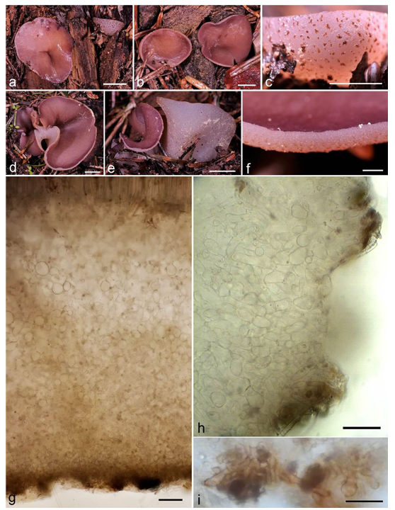
Fig. 44 Hansenopezia decora. a-f Ascomata. g Vertical median section through excipular middle flank. h Marginal tissue. i Terminal cells of excipular pustule. g–i In water mount (*). a–c, g, i From CNF 2/10621 (holotype), d–f From CNF 2/10618, h From CNF 2/7823. Photo: N. Matočec and I. Kušan. Scale bars: a, b, d, e = 1 cm, c = 0.5 cm, f = 0.1 cm, g = 100 µm, h, i = 50 µm. Asterisk (*) denotes living material

Fig. 45 Hansenopezia decora. a Contact zone of subhymenium and medullary excipulum. b Medullary excipulum. c Ectal excipulum with a pustule. d Living asci with ascospores. e Ascus bases with ascogenous cells. f Paraphyses. g Amyloidity of ascal mucus. h Ascospores. a–f, h In water mount (*), g In Lugol's solution (*/†). a,b phase contrast, c–h bright field. All from CNF 2/10621 (holotype). Photo: N. Matočec and I. Kušan. Scale bars: a–c = 50 µm, d–h = 10 µm. Asterisk (*) denotes living material. Cross (†) denotes dead material