Ganoderma eghatensis Kezo, K., Gunaseelan, S., & Kaliyaperumal, M., sp. nov
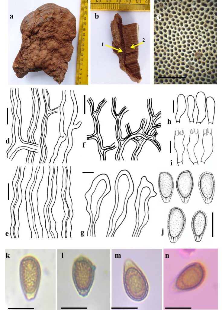
MycoBank number: MB 557014; Index Fungorum number: IF 557014; Facesoffungi number: FoF 10728; Fig. 2
Etymology: “eghatensis” refers to the place of collection.
Holotype: MUBL4014
Description: Basidiocarps annual, solitary to cluster, sessile, applanate, leathery when fresh and woody to corky when dry. Pileus semicircular, convex, projecting up to 10 cm, 9 cm wide and 4 cm thick near the base. Pilear surface reddish brown (9E8) when fresh, liver brown (7D6) when dry, covered by a laccate layer, hard and irregular surface towards the attachment becoming concentrically zonate near the margin. Margin acute, white (7A1) when fresh, orange white (6A2) when dry. Pore surface orange white (6A2) to brownish orange (6C7). Pores round regular, 4–8 per mm. Context up to 1.5 cm, homogeneous, light brown (6D6) to brown (7E8). Tubes brown (6D6) to light brown (6D6), up to 3 cm thick.
Hyphal system trimitic, Skeletal hyphae dominant in the basidiomata. Context Generative hyphae, thin to thick walled, hyaline, clamped septate, branched, 2.5 – 6 μm diam.; skeletal hyphae, thick-walled with narrow lumen, unbranched, rust brown, aseptate, 3.7 – 8 μm diam.; binding hyphae, thick-walled with narrow lumen, branched, aseptate, 2.5 – 4 μm diam. Trama Generative hyphae, thin to thick walled, hyaline, rarely clamped septate, frequently branched, 2 –5 μm diam.; skeletal hyphae, thick walled with narrow lumen, rust brown, aseptate, unbranched, 3.7 – 7.5 μm diam.; binding hyphae, thick-walled with narrow lumen, branched, aseptate, 2.5 – 4.7 μm diam. Cutis cell broadly clavate, rarely umbonate, 27.5 – 60 × 10 – 18.7 μm. Cystidioles absent. Basidioles clavate, 15.5 – 25 × 5 – 6.2 μm. Basidia clavate, with four sterigmata, 17 – 30 × 5 – 8.5 μm. Basidiospores ellipsoid, brown, thick-walled, truncate at the apex, bitunicate, exospore thin, subhyaline, smooth, endospore thick, brown, echinulate; (11.2-) 11.5 – 14.7 (-15) × (7.5-) 7.7 – 9 (-9.5) μm (n = 50/2), Q = 1.3 – 1.5; CB ̄, IKI ̄.
Specimen examined: INDIA, Tamil Nadu, Thiruvannamalai district, Jawadhu hills, Nallapathur Reserve Forest, 12°32’49.9″N 78°54’05.9″E, on dead wood, 14 January 2020, Kezhocuyi Kezo, (Holotype, MUBL4014).
Additional specimen examined: INDIA, Tamil Nadu, Salem district, Yercaud, 11°48’12.4″N 78°16’03.0″E, on dead wood, 26 February 2020, Kezhocuyi Kezo (Isotype, KSM-AR1). INDIA, Tamil Nadu, Salem district, Yercaud, 11°48’15.1″N 78°16’13.6″E on dead wood, 26 February 2020, Kezhocuyi Kezo (Isotype, KSM-AR4). INDIA, Tamil Nadu, Salem district, Yercaud, 11°49’01.8″N 78°15’37.4″E, on dead wood, 26 February 2020, Kezhocuyi Kezo (Isotype, KSM-VR6). INDIA, Tamil Nadu, Salem district, Kolli Hills, 11°19’46.0″N 78°21’22.8″E, on dead wood, 26 February 2020, Kezhocuyi Kezo (Isotype, KSM-KV3). INDIA, Tamil Nadu, Thiruvannamalai district, Jawadhu hills, 12°28’58.0″N 78°49’26.7″E, on dead wood, 14 January 2020, Kezhocuyi Kezo (Isotype, KSM-TM61).
GenBank numbers: ITS: MZ203592, LSU- OM432453, β-tubulin: MZ229331
Notes: Ganoderma eghatensis shares similarity with G. mizoramense by having semicircular and convex pileus, presence of laccate pilear layer, acute margin, absences of cystidioles and reddish brown basidiocarp but the former differs by smaller basidiocarp size and uniform colour of upper margin and pilear surface; smaller pore and larger pore range (4-8/mm), larger spore size (11.2-) 11.5 – 14.7 (-15) × (7.5-) 7.7 – 9 (-9.5) μm (Crous et al., 2017; Zohmangaiha et al., 2019). G. eghatensis differs in having larger pores and basidiospores other closely-related species namely G. destructans, G. martinicense, G. mizoramense, G. multiplicatum, G. multipileum and G. steyaertanum (Hapuarachchi et al. 2015; Cabarroi-Hernández et al., 2019; Tchotet Tchoumi et al., 2019) found in Cluster A.3, Clade A of Ganoderma spp. meta-analysis as represented by Fryssouli et al. (2020).

Fig. 1: Ganoderma eghatensis (MUBL4014, halotype). a Basidiocarp. b1 Context. b2 tube layer. c Pore surface. d Generative hyphae. e Skeletal hyphae. f Binding hyphae. g Cutis cell. h Basidioles i Basidia. j Basidiospores. k Basidiospore in H2O. l Bsidiospore in cotton. m Basidiospore Melzer’s reagent. blue. n Basidiospore in phloxine. Scale: c = 1 mm d – n = 10 µm.
