Fusoidigranularius W. Dong, H. Zhang & K.D. Hyde, gen. nov.
Index Fungorum number: IF557491; Facesoffungi number: FoF 09544
Etymology: – in reference to its fusoid ascospores with a large granular sheath
Saprobic on submerged wood.
Sexual morph: Ascomata solitary or gregarious, immersed, obpyriform, oriented horizontally to the host substrate, black, coriaceous, with ostiolate papilla, with an upward bend, relatively long neck growing laterally, periphysate. Peridium comprising several layers of thick-walled, flattened cells of textura angularis, outer layer dark brown and encrusted with pigmented particles, hyaline and unencrusted inwardly. Paraphyses rarely septate, hyaline, unbranched, tapering distally. Asci 8-spored, unitunicate, cylindrical, long pedicellate, persistent, with a large, refractive, J-, apical ring. Ascospores uniseriate to overlapping uniseriate, fusoid, 5–9(–11)-septate, hyaline, smooth-walled, with a large, gelatinous, irregular, granular sheath.
Asexual morph: Undetermined.
Type species: – Fusoidigranularius nilensis (Abdel-Wahab & Abdel-Aziz) W. Dong, H. Zhang & K.D. Hyde
Notes: – Annulatascus nilensis was collected from submerged stems of Phragmites australis
in the river Nile in Egypt (Abdel-Wahab et al. 2011). It clustered with Annulatascus species with weak bootstrap support, and was placed in Annulatascus based on limited data (Abdel-Wahab et al. 2011). Annulatascus nilensis appears to have typical morphological characteristics of Annulatascus, such as cylindrical asci, long pedicel, and a large, refractive apical ring (Abdel-Wahab et al. 2011). However, A. nilensis has immersed ascomata that oriented horizontally to the host substrate and with an upwardly bending neck growing laterally, which are distinctive from Annulatascus, and even from Annulatascaceae. In addition, A. nilensis has 5–9(–11)-septate ascospores with an irregular, granular sheath that differ from Annulatascus species, including our three new species. Phylogenetically, A. nilensis forms a separated clade from Annulatascus in our updated phylogenetic tree (Fig. 1), which corroborates other studies (Hyde et al. 2018, 2020, Luo et al. 2019). The new genus Fusoidigranularius is, therefore, established to accommodate A. nilensis based on distinct morphology and phylogenetic analyses.
The ascomata lying horizontally is the key feature of two annulatascaceae-like families, Atractosporaceae and Pseudoproboscisporaceae. However, the ascomata of Atractosporaceae and Pseudoproboscisporaceae are semi-immersed to nearly superficial and they have aseptate or few septate ascospores (Zhang et al. 2017). A new collection of Fluminicola saprophytica (MFLUCC 18-1244) found in this study also has immersed ascomata that oriented horizontally to the host substrate and with an upward bend neck growing laterally (Fig. 26), which are similar to Fusoidigranularius. However, they can be distinguished by septation and shape of ascospores. Although Fusoidigranularius has a weak relationship with Annulatascaceae members (Fig. 1), it is temporarily placed in Annulatascaceae based on current phylogenetic analysis.
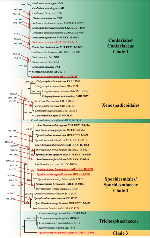
Figure 1 – RAxML tree generated from combined LSU, ITS, TEF and RPB2 sequence data. Bootstrap support values for maximum likelihood (the first value) equal to or greater than 75% and Bayesian posterior probabilities (the second value) equal to or greater than 0.95 are placed near the branches as ML/BYPP. The tree is rooted to Aureobasidium pullulans CBS 584.75 and Dothidea insculpta CBS 189.58. The ex-type strains are indicated in bold and newly generated sequences are indicated in red. The new species introduced in this study are underlined.
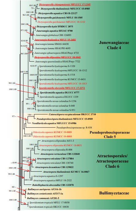
Figure 1 – Continued.
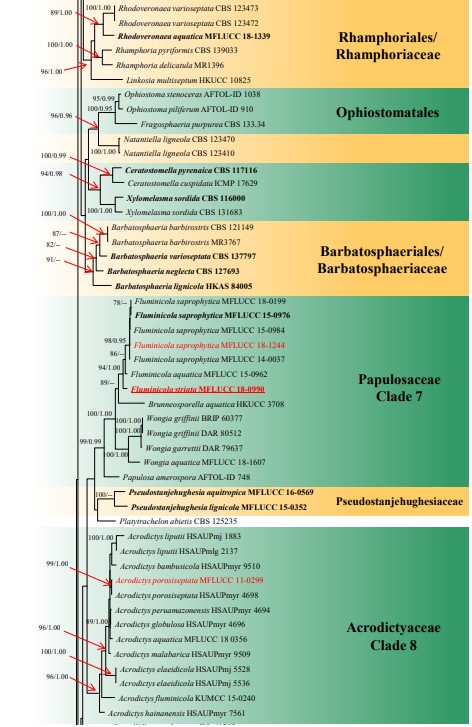
Figure 1 – Continued.
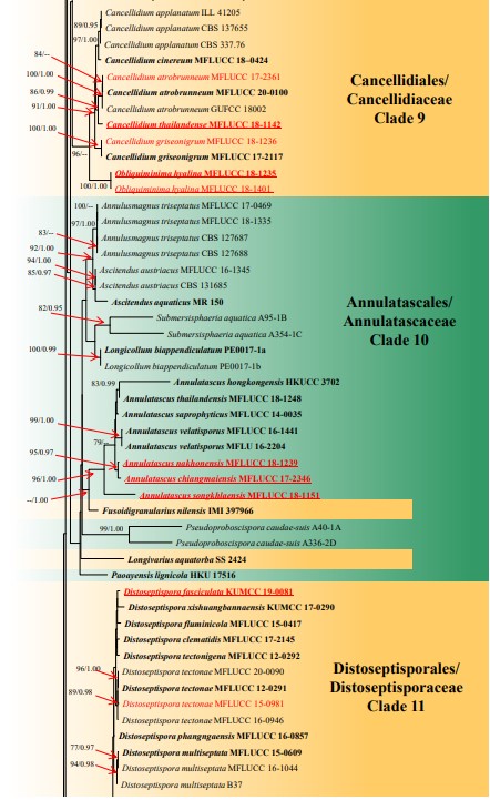
Figure 1 – Continued.
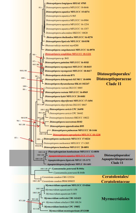
Figure 1 – Continued.
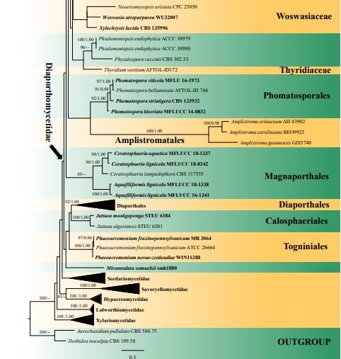
Figure 1 – Continued.
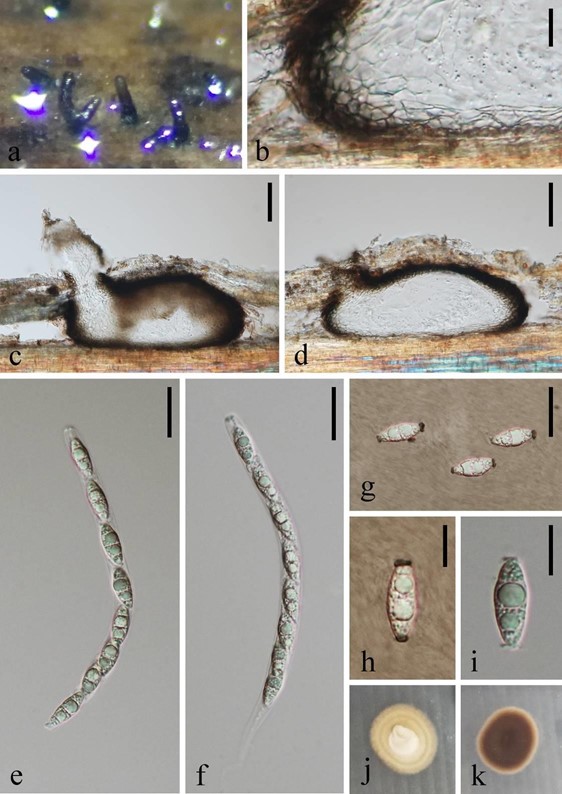
Figure 26 – Fluminicola saprophytica (MFLU 18-1567). a Appearance of necks of ascomata on host. b Structure of peridium. c, d Vertical sections of ascomata. e, f Unitunicate asci. g, h Ascospores in Indian Ink. i Ascospore in water. j, k Colony on PDA (left-front, right-reverse). Scale bars: b, h, i = 10 μm, c, d = 50 μm, e–g = 20 μm.
Species
