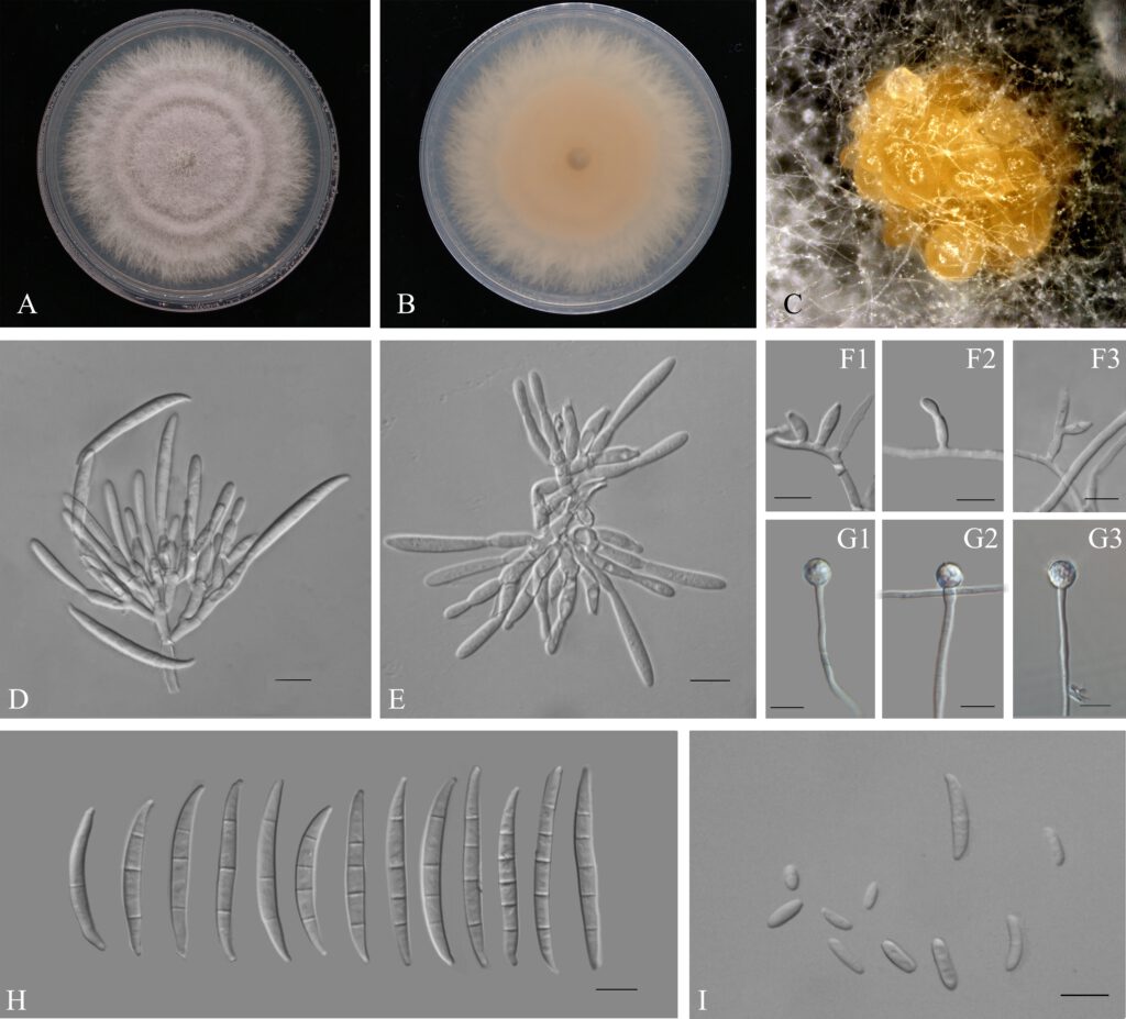
Fusarium elaeidis L. Lombard & Crous, in Lombard, Sandoval-Denis, Lamprecht & Crous, Persoonia 41: 23 (2018) (Figure 1)
MycoBank number: MB 826838; Index Fungorum number: IF 826838; Facesoffungi number: FoF 10248;
Micromorphology: Pathogenic on Aglaonema modestum. Sexual morph: Not observed. Asexual morph: Sporodochia pale orange, formed abundantly on carnation leaves. Sporodochial conidiogenous cells 7–20 × 2–4 µm (x̅ = 12 × 3 µm n = 50), doliiform to subcylindrical, smooth and thin-walled, with periclinal thickening and an inconspicuous apical collarette. Sporodochial macroconidia falcate, curved dorsiventrally with almost parallel sides tapering slightly towards both ends, with a blunt to papillate, curved apical cell and a blunt to foot-like basal cell, 3–5-septate, hyaline, smooth- and thin-walled; 3-septate conidia: 30–50 × 3–5(–6) µm (x̅ = 38 × 4 µm, n = 50); 4-septate conidia: 35–55(–60) × 3–5(–6) µm (x̅ = 45 × 4 µm, n = 50); 5-septate conidia: 40–50(–55) × 4–5 µm (x̅ = 47 × 4 µm, n = 15). Conidiogenous cells 5–15 × 2–4 µm (x̅ = 9 × 3 µm, n = 50), subulate, to subcylindrical, smooth- and thin-walled; aerial microconidia forming small false heads on the tips of the phialides, 0–1-septate; 0-septate conidia: 5–15(–20) × 2–3 µm (x̅ = 8 × 3 µm n = 50); 1-septate conidia: 10–25 × 2–4 µm (x̅ = 18 × 3 µm, n = 16). Chlamydospores 5–10 × 5–10 µm (x̅ = 9 × 8 µm), formed solitary, in pairs or chains, either terminal or intercalary in hyphae.
Culture characteristics: Colonial growth rate was 5.1 mm on PDA at 25 ℃ per day. The colony surface was light purple, flat, and floccose with a regular margin. In reverse colony white at first and turned rosy vinaceous in the centre at the end. On CLA, aerial mycelium sparse with abundant orange sporodochia forming on the carnation leaves.
Material examined: China, Guangdong province, Guangzhou city, Aglaonema modestum Schott ex Engl., (Araceae). 31st March 2021, Y.X. Zhang (dried cultures ZHKU 220047, ZHKU 220048 and ZHKU 220049); living cultures, ZHKUCC 220080, ZHKUCC 220081 and ZHKUC220082.
Notes: In the phylogenetic tree, three isolates in this study clustered with Fusarium elaeidis with 90% ML and 0.99 BYPP. Morphologically the length of microconidia of our isolates is longer than that of microconidia as described by Lombard et al. (2019). The base pairs of both tef 1-α and β-tubulin in this study (ZHKUCC 220047) are the same as that of F. elaeidis (CBS 217.49). Only one base pairs of rpb2 (totally 616 bp) were different from that of F. elaeidis (CBS 217.49) in Lombard et al. study (2019). Therefore, based on phylogenetic analyses and morphological analyses, our isolates from A. modestum were identified as F. elaeidis.

Fig. 1| Fusarium elaeidis. (A–B) Culture characteristics on PDA after 7 days (A, upper view; B, reverse view). (C) Sporodochia formed on the surface of carnation leaves. (D–E) Sporodochial conidiophores and phialides (F1–F3) Aerial conidiophores and phialides. (G1–G3) Chlamydospores. (H) Sporodochial conidia. (I) Aerial conidia. Scale bars: (D–I) = 10 μm.a
