Erysiphe heraclei DC., Fl. franç., Edn 3 (Paris) 5/6: 107 (1815)
Index Fungorum number: IF 120607
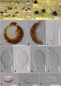
Saprobic on living stem of Torilis arvensis. Asexual morph: Undetermined. Sexual morph: Ascomata 70–80 × 85–90 µm (x̄ = 76 × 87 µm, n = 10), immersed to semi-immersed, solitary to aggregate, sessile, globose, uniloculate. Peridium 20–28 µm (x̄ = 23 µm, n = 10), slightly wide at the sides, comprising of 3–4 cell layers, outer most layer comprising heavily pigmented, thick-walled, dark brown to pale brown cells of textura angularis, inner layer comprising pale brown to hyaline cells of textura angularis. Hamathecium comprising numerous, 2–4 µm wide (x̄ = 2.8 µm, n = 10), filamentous, aseptate, pseudoparaphyses. Asci 50–80 × 35–46 µm (x̄ = 58 × 39 µm, n = 10), 4-spored, bitunicate, saccate or broadly obpyriform, straight or slightly curved, apically rounded with indistinct ocular chamber, short pedicellate. Ascospores 14–21 × 11–15 µm (x̄ = 17 × 13 µm, n = 20), globose to subglobose, aseptate, with rounded ends, hyaline in both immature and mature stages, smooth-walled.
Material examined: Italy, Forlì-Cesena Province, a living stem of Torilis arvensis (Apiaceae), 6 August 2018, Erio Camporesi, IT3973 (MFLU 18-1489).
GenBank numbers: ITS: MT921775, LSU: MT921669.
Known distribution (based on molecular data): Azerbaijan, Tengaltı Province (Abasova et al. 2018a), Italy, Forlì-Cesena Province (this study).
Known hosts (based on molecular data): Conium maculatum (Qiu et al. 2019), Torilis arvensis (Abasova et al. 2018a), this study).
Notes: – All powdery mildew species are exclusively obligate biotrophs of plants, i.e., they feed on living plants. Ascospores did not germinate in PDA and MEA media at different temperatures. So, DNA was obtained directly from the fruiting bodies. Erysiphe heraclei is most common on Apiaceae hosts worldwide (Braun & Cook 2012). In this study, we introduce the first geographical record of Erysiphe heraclei in Italy.

Fig. 1 – Erysiphe heraclei (MFLU 18-1489, new geographical record). a, b Appearance of ascomata on a living stem of Torilis arvensis. c Longitudinal section of ascomata. d Peridium. e Pseudoparaphyses. f–i Asci. j–l Ascospores. Scale bars: a = 200 μm, b = 100 μm, c, e = 50 μm, d, f–i, l = 20 μm, j–k = 10 μm.
