Eriocamporesia aurantia R.H. Perera, Samarak. & K.D. Hyde, sp. nov.
MycoBank number: MB 556792; Index Fungorum number: IF 556792; Facesoffungi number: FoF 06960; Figs. 106, 107
Etymology: Name reflects orange stromata.
Holotype: MFLU 19-0943
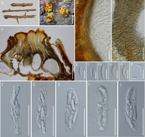
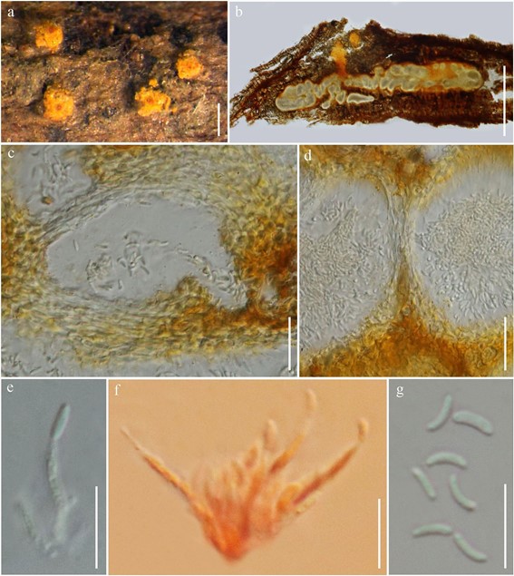
Associated with twigs of dicotyledonous plant. Sexual morph Ascostromata on host single, appearing as yellow pustules, 450–670 μm high, 870–1000 μm diam., pulvi- nate, semi-immersed in plant tissue, orange, KOH + purple; ascostromatic tissue pseudoparenchymatous, covering the top of the ascomatal bases. Ascomata 450–700 μm high, 120–330 μm diam., perithecial, usually valsoid, embedded in stromata at irregular levels, fuscous black, necks emerge at stromatal surface as dark orange brown ostioles covered with orange stromatal tissue to form papillae extending above stromatal surface. Peridium 12–24 μm wide, composed of 5–7 layers of flattened, elongate, brown-walled, cells of textura angularis. Paraphyses lacking. Asci 30–43 × 6.7–8.7 µm ( x̄ = 37.6 × 7.8, n = 14), 8-spored, unitunicate, oblong ellipsoidal to fusoid to ellipsoidal, apedicellate, floating freely in the perithecial cavity, thin-walled, apex simple. Ascospores 9.5–14.5 × 2.5–3.3 μm ( x̄ = 11.6 × 2.8, n = 30), 2-seriate, cylindrical to allantoid, occasionally ellipsoidal, ends round, curved or sometimes straight, aseptate, hyaline, smooth-walled, without appendages. Asexual morph Conidiostromata 445–630 μm high, 1500–1800 μm diam., appearing as yellow pustules on the host, occur as separate structures, pulvinate, semi-immersed, orange. Conidiomata multi-locular, with locules often convoluted, apapillate, with 1-ostiolar opening, orange, KOH + purple; stromatic tissue pseudoparenchymatous. Paraphyses lacking. Pycnidial walls 4–7 μm wide, composed of 2–4 layers of flattened, elongate, hyaline, cells of textura angularis. Conidiophores 16.3–23.6 μm long, 1.6–2.3 wide, cylindrical, rarely septate or branching, hyaline. Conidiogenous cells 8.8–9.7 μm long, 1.1–1.6 wide, phialidic, cylindrical with attenuated apices, hyaline. Conidia 3.7–5.7 × 1–1.5 μm (x̄ = 4.9 × 1.2, n = 30), hyaline, cylindrical or allantoid, ends round, aseptate, hyaline, smooth-walled, without appendages.
Culture characteristics: Ascospores germinated on PDA within 12 h, colonies on PDA reaching 35–38 mm diam. after 2 weeks at 25 °C, mycelium partly superficial, partly immersed, slightly effuse, with irregular edge, initially white, becoming orangish with time.
Material examined: THAILAND, Chiang Mai Province, Mae Rim District, on twigs of dicotyledonous plant, R.H. Perera, 9 September 2017, Rim19 (MFLU 19-0943, holo- type), ex-type living culture, MFLUCC 17-2625.
GenBank numbers: ITS =MN699135, LSU =MN699130.
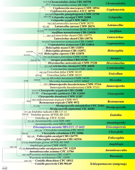
Notes: Eriocamporesia aurantia clusters within Cryphonectriaceae as a separate lineage (Fig. 108). BLASTn search of the ITS rDNA region of our new isolate showed the highest similarity to Amphilogia gyrosa (Berk. & Broome) Gryzenh., H.F. Glen & M.J. Wingf. strain 91123101 (94% similarity, 465/494 bp), Endothia gyrosa (Berk. & Broome) Höhn. strain AFTOL-ID 1223 (94% similarity, 531/564 bp),mE. radicalis (Schwein. ex Fr.) Tul. & C. Tul. strain CBS 116.13 (94% similarity, 549/585 bp), Chrysoporthe inopina Gryzenh. & M.J. Wingf. strain CMW 12731 (94% similarity, 549/585 bp) and strain CBS 116.13 (94% similarity, 543/580 bp). Eriocamporesia aurantia shares similar asexual morph characters to Amphilogia (Gryzenhout et al.2005a; Senanayake et al. 2017). However, it can be distinguished from Amphilogia species in having valsoid perithecia, and aseptate, cylindrical to allantoid ascospores, while Amphilogia has diatrypoid perithecia, and 1–3-septate, fusoid to ellipsoid ascospores (Gryzenhout et al. 2005a; Senanayake et al. 2017). Considering both molecular and morphological data, we introduce E. aurantia as a novel species.

Fig. 106 Sexual morph of Eriocamporesia aurantia (MFLU 19-0943, holotype). a Herbarium material. b Ascostromata on host. c Longitudinal section of ascostroma. d Section of peridium. e Peridium in face view. f–j Asci. k–o Ascospores. p Germinating ascospore. Scale bars: b, c = 200 μm, d–j = 20 μm, k–o = 10 μm, p = 20 μm

Fig. 107 Asexual morph of Eriocamporesia aurantia (MFLU 19-0943, holotype). a Conidiostromata on host. b Longitudinal section of conidiostroma. c Section of conidioma. d Section of peridium. e, f Conidiophores with conidia (f in Congo red). g Conidia. Scale bars: a = 100 μm, b = 500 μm, c, d = 20 μm, e–g = 10 μm

Fig. 108 Phylogram generated from maximum likelihood analysis based on combined LSU, ITS and TUB2 sequence data representing Cryphonectriaceae. Related sequences are taken from Senanayake et al. (2017). Fifty-four strains are included in the combined analyses which comprise 1830 characters (866 characters for LSU, 639 char- acters for ITS, 323 characters for TUB2) after alignment. Coniella tibouchinae (CPC 18512) and C. quercicola (CBS 904.69) in Schizoparmaceae is used as the outgroup taxa. Single gene analyses were also performed to compare the topology and clade stability with com- bined gene analyses. Tree topology of the maximum likelihood analy- sis is similar to the Bayesian analysis. The best RAxML tree with a final likelihood values of − 7862.571574 is presented. The matrix had 525 distinct alignment patterns, with 29.64% undetermined characters or gaps. Estimated base frequencies were as follows: A = 0.239368, C = 0.254757, G = 0.280128, T = 0.225747; substitution rates AC = 2.087607, AG = 3.025115, AT = 2.060161, CG = 1.371584, CT = 7.165898, GT = 1.000000; gamma distribution shape parameter α = 0.727546. Bootstrap values for maximum likelihood (ML) equal to or greater than 50% and clade credibility values greater than 0.90 (the rounding of values to 2 decimal proportions) from Bayesian- inference analysis labelled on the nodes. Ex-type strains are in bold and black, the new isolate is indicated in bold and blue
