Epicoccum viciae-villosae W. Zhao, Q. Ning, & J.Y. Yan, sp. nov
MycoBank number: MB 558424; Index Fungorum number: IF 558424; Facesoffungi number: FoF 10794;
Description
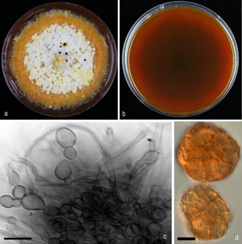
Asexual morph: Conidiomata sporodochial, solitary or aggregated, immersed to semi-immersed, glabrous, covered with hyphal growth, blackish brown. Hyphae smooth, branched, septate, hyaline. Chlamydospores multicellular, produced in agar, hyaline to pale brown, subglobose or oblong, 10–20×6–9.5 μm(x̅ =16.3×8.1 μm,n = 10). Conidia, multicellular, yellowish-brown to brown, globose to subglobose, 25–67.5×20–51.5 μm(x̅ =37.3×29.7 μm,n = 40). Sexual morph: not observed.
Cultural characteristics – Colonies on PDA are slow-growing, covering a 30 mm Petri dish in 10-14 days after incubation at 25 ± 1 °C, yellowish white (surface) and reddish black (reverse), with a dense mat of aerial mycelium, rough, entire slightly radiating at the margin; colony from above rough, white at fruiting zone, slimy reddish spore mass at the centre, and yellowish at ageing zone. Later give reddish-brown colour to the PDA media.
Material examined: China, Henan Province, Shihe District, from Vicia villosa pod, May 2018, Zhao Wensheng and Zhang Guozhen (JZBH 380079 holotype); ex-type living culture = JZB 380079.
Additional material examined – China, Henan Province, Shihe District, from Vicia villosa pod, May 2018, Zhao Wensheng and Zhang Guozhen, living culture JZB 380080, JZB 380081, JZB 380082, JZB 380086, Henan Province, Shihe District, from Vicia villosa stem, May 2018, Zhao Wensheng and Zhang Guozhen, living culture JZB 380083, Henan Province, Shihe District, from Vicia villosa leaf, May 2018, Zhao Wensheng and Zhang Guozhen, living culture JZB 380084, Henan Province, Shihe District, from Vicia villosa stem, May 2018, Zhao Wensheng and Zhang Guozhen, living culture JZB 380058, JZB 380059.
Distribution: China
Notes: In multi-loci phylogenetic analysis of Epicoccum species using LSU, ITS, rpb2, and tub markers, Epicoccum viciae-villosae developed a monophyletic clade with 84% ML support (Fig. 5) with E. layuense. Epicoccum viciae-villosae has relatively larger conidia than E. layuense (25.4–67.5×20.1–51.5 μm vs 13–19.5 μm). Furthermore, E. layuense produced subglobose -pyriform conidia, with a basal cell, that we could not find in Epicoccum viciae-villosae.

Fig. x. Epicoccum viciae-villosae a, b Colony on PDA, 10 days after incubation at 25 ± 1 °C (a from above, b from below) c Sporodochia d Conidia. Scale bars: c, d= 10 µm.
