Entodesmium artemisiae S. Konta, Bulgakov & K.D. Hyde.
Index Fungorum number: IF551450; Facesoffungi number: FoF00950; Fig. 1
Etymology: The specific epithet refers to the host genus Artemisia.
Holotype – MFLU 15-0007
Saprobic on on dead stems of Artemisia. Sexual morph: Ascomata 205 – 292 μm wide, 177 – 258 μm high (x̄ = 231.3 μm × 207 μm), scattered, solitary, dark brown, semiimmersed to erumpent, subglobose, ostiolate. Ostiole lined with hyaline periphyses. Peridium 26 – 41 μm wide (x̄ = 35.08 μm), comprising two strata, outer stratum comprising black, thick – walled, occluded cells, inner stratum comprising 3 – 4 layers of black, thick – walled cells of textura angularis. Hamathecium comprising numerous, 1.5 – 2.5 μm wide (x̄ = 2 μm), filamentous, branched, septate pseudoparaphyses. Asci 80.5 – 112.5 × 9 – 14 μm (x̄ = 95.6 × 11.22 μm, n = 20), 8 – spored, bitunicate, cylindric – clavate, pedicellate, apically rounded, with an ocular chamber. Ascospores 61.5 – 96.5 × 2.5 – 4 μm (x̄ = 82.96 × 2.13 μm, n = 40), fasciculate, initially hyaline, becoming yellowish brown to brown at maturity, filiform, slightly curved, widest and curved approximately 1/3rd from apex, 13 – 20 – septate, lacking a sheath or appendages, not breaking into part spores. Asexual morph: Undetermined.
Culture characteristics – Colonies on MEA, reaching 5.5 cm diam. after 2 weeks at 16 °C, olive brown in the middle, yellowish white at the edges, and reverse olive brown at the margins, medium dense, irregular, flattened to slightly raised, with rough surface, crenated radiating margins, floccose to fairly fluffy, brown pigment produced in agar.
Material examined – RUSSIA, Rostov region, Shakhty City, Salty Hollow, stony steppe, dead stems of Artemisia campestris L., 3 June 2014, T. Bulgakov, T109 (MFLU 15-0007, holotype); ex-type living culture, MFLUCC 14-1156.
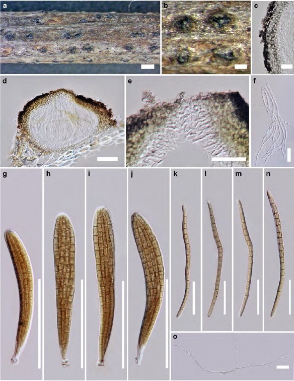
Fig. 1 Entodesmium artemisiae (holotype) a Appearance of ascomata on host substrate b Close up of ascomata which are erumpent through host epidermis c Cell arrangement of peridium d Section of ascoma e Periphyses f Pseudoparaphyses g – j Asci k – n Ascospores o Germinating ascospore. Scale bars: a = 500 μm, b = 200 μm, c = 20 μm, d = 50 μm, e – f = 20 μm, g – j = 50 μm, k – n = 20 μm, o = 50 μm.