Elongatopedicellata lignicola J.F. Zhang, J.K. Liu, K.D. Hyde & Z.Y. Liu, sp. nov.
Index Fungorum number: IF551485; Facesoffungi number: FoF00960 Fig. 1
Etymology – from the Latin lignin and cola, in reference to its habit on wood.
Holotype – MFLU 15-2717
Saprobic on the dead wood. Sexual morph: Ascomata 294 – 387 μm high (including neck), 187 – 225 μm diam., solitary to gregarious, scattered, immersed or erumpent, uni – loculate, subglobose to obpyriform, coriaceous, visible on host surface as raised, dark spots, with papillate ostiole. Ostiole central, with pore – like opening and papilla to long neck. Peridium 14 – 21 μm wide, of unequal thickness, slightly thin at the base, composed of several layers of brown to dark brown, thick-walled cells, arranged in a textura angularis. Hamathecium composed of 1 – 2 μm wide, filiform pseudoparaphyses, anastomosing between and above the asci, embedded in a gelatinous matrix . Asci ( 59.5 – ) 63 – 132(−137) × (9.5–) 10 – 12.5 (−13.5) μm (x̄ = 98.7 × 12.2 μm, n = 25), 8 – spored, bitunicate, fissitunicate, fusiform-clavate, with a long foot-like or knob – like pedicel, apically rounded, with a well – developed ocular chamber. Ascospores (18.5–) 19 – 21 (−22) ×( 4.9) – 5.2 – 6 (−6.3) μm (x̄ = 20.3 × 5.8 μm, n = 30), 1 – 3 overlapping seriate, hyaline, fusiform with narrow ends, 1 – septate, constricted at the septum, upper cell shorter and wider, lower cell long and narrow, slightly curved, surrounded by a mucilaginous sheath. Asexual morph: Undetermined.
Material examined − THAILAND, Chiang Rai, Muang District, Mae Chang Hot Spring, on dead branch, 25 November, 2014, JF Zhang (MFLU 15-2717, holotype); ex-type living culture, MFLUCC 15-0642, CFTCC.
Culture characteristics − Colonies on PDA fast growing, reaching 35 – 38 mm diam. after 2 weeks at 25 °C, cream to pale gray at the margin, buff pigment produced at the center, reverse cream to pale yellowish white at the margin, yellowish to pale reddish at the center, slighting radiating, medium dense, raised, dull with undulate edge, fairy fluffy at the margins, separating from agar.
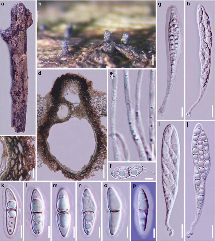
Fig. 1 Elongatopedicellata lignicola (holotype) a, b Appearance of ascomata on host surface c Section of peridium d Vertical section through ascoma e Pseudoparaphyses f Germinating spore g – j Asci k – o Ascospores p Ascospore in Indian ink showing sheath. Scale bars: b = 200 μm, c, h, j = 10 μm, d = 50 μm, e – g, i, k – p = 5 μm.