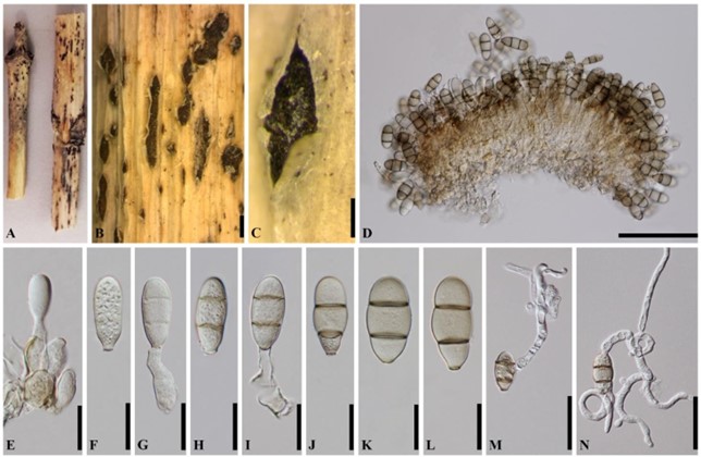
Dothidotthia negundinicola (Crous & Akulov) Senwanna, Wanas., Bulgakov, Phookamsak & K.D. Hyde, Mycosphere 10(1): 716 (2019). Fig. 80
≡ Neodothidotthia negundinicola Crous & Akulov, in Crouset al., Fungal Systematics and Evolution 3: 93 (2019).
MycoBank number: MB 556640; Index Fungorum number: IF 556640; Facesoffungi number: FoF 06139.
Associated with canker on twigs of Acer negundo (Sapindaceae). Sexual morph: Undetermined. Asexual morph: Colonies 180–470 µm diam, partly immersed, ascostromatic, effuse, sporodochial, with partly immersed, basal pseudoparenchymatous ascostroma, erumpent, black, velvety. Conidiophores (16–)21–33(–36) × 5–11 µm ( x̅ = 27.6 × 8.5 μm, n = 20), semi- macronematous, septate, branched, subhyaline, smooth, arising from basal ascostroma. Conidiogenous cells 13–26 µm long, monoblastic, integrated, terminal. Conidia (24–)28–36(–38) × 10–16.5 µm (x̅ = 32.2 × 16.5 μm, n = 75), acrogenous, fusiform to obclavate to obpyriform, pale to brown, truncate at base, with a protruding hilum, rounded at apex, 2-septate, constricted at septa, minutely echinulate.
Culture characteristics – Colonies on MEA, reaching 3 cm diameter after 2 weeks at 25– 30 °C, producing dense mycelium, circular, velvety to woolly, rough margin, white to creamy-grey, with aerial mycelium.
Material examined – Russia, Rostov region, Shakhty Park, on dead and dying twig of Acer negundo (Sapindaceae), 1 March 2016, Timur S. Bulgakov, T-1494 (MFLU 16-1788), living culture MFLUCC 17-2511.
GenBank numbers – ITS: MN168763, LSU: MN168760, SSU: MN168758.
Notes – Crous et al. (2019b) reported and illustrated Neodothidotthia negundinicola (CBS 145039) from Acer negundo in Ukraine. Senwanna et al. (2019) synonymized Neodothidotthia negundinicola under Dothidotthia negundinicola based on morphology and phylogeny. The conidial morphology of our fresh specimen resembles Dothidotthia negundinicola (CBS 145039) in having fusiform to obclavate, pale to brown, 2-septate, 24–38 × 10–16.5 µm and in the combined multi-gene phylogeny.

Figure 80 – Dothidotthia negundinicola (MFLU 16-1759). a–c Sporodochia on host surface. d Vertical section of sporodochium. e Conidia attached with the conidiogenous cells. h–l Conidia. m, n Germinated conidia. Scale bars: b = 500 µm, c = 200 µm, d = 100 µm, e–l = 20 µm, m, n = 40 µm.
