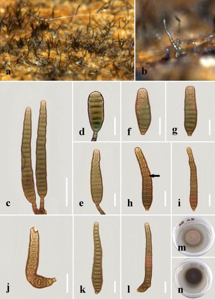
Distoseptispora pachyconidia R.Zhu & H. Zhang, sp. nov.
MycoBank number: MB; Index Fungorum number: IF; Facesoffungi number: FoF 12581;
Description
Saprobic on decaying wood submerged in freshwater. Sexual morph: undetermined. Asexual morph: hyphomycetous. Colonies on the substratum superficial, effuse, hairy or velvety, dark-brown or black. Mycelium mostly immersed, consisting of branched, septate, smooth, hyaline to pale-brown hyphae. Conidiophores 11–27 μm long (x̄=19 µm, n=10), 4–9 μm wide (x̄=6 µm, n=10), macronematous, mononematous, solitary, unbranched, 2–4-septate, cylindrical, straight or flexuous, smooth, pale-brown to mid-brown, slightly tapering distally, truncate at the apex. Conidiogenous cells 5–9 μm long (x̄=7 µm, n=10), 4–6.5 μm wide (x̄=5 µm, n=10), monoblastic, integrated, determinate, terminal, cylindrical, pale-brown, smooth. Conidia 42–136 μm long (x̄=84 μm, n=30), 14–22 μm at the widest part (x̄=17 μm, n=30), 8–14 μm wide at the apex (x̄=11 μm, n=30), acrogenous, solitary, obclavate, lanceolate, rostrate or not, straight or curved, 8–21-distoseptate, truncate at the base, tapering towards the rounded apex, smooth, thick-walled, pale-brown with a green tinge.
Material examined: China, Yunnan Province, Xishuangbanna, Na Dameng Reservoir, found on dead, submerged, decaying wood of unidentified plants, 7 November 2020, Rong Zhu, N37 (HKAS 122179, holotype), ex-type living culture KUNCC 21–10724.
Distribution: China
Sequence data: ITS: OK310696 (ITS5/ITS4); LSU: OK341194 (LROR/LR5); EF1a: OP413477 (983/2218R); RPB2: OP413471 (fRPB2-5F/fRPB2-7cR)
Notes: Distoseptispora pachyconidia clusters as an independent branch in Distoseptispora based on a concatenated LSU–ITS–TEF1-α–RPB2 phylogeny. It is phylogenetically close to D. crassispora, D. chinense and D. tectonigena. Compared to D. tectonigena, D. pachyconidia has shorter conidiophores (11–27 μm vs. up to 110 μm) and shorter conidia (42–136 μm vs. 148–225(–360) μm), as well as fewer conidial septa (8–21 vs. 20–46) [3]. In addition, the conidia of D. tectonigena were sometimes percurrently proliferating 5–10 times at the apex, which was not observed in D. pachyconidia. The conidia in D. pachyconidia are light-colored and shorter than those in D. crassispora and D. chinense (42–136 μm vs. 95–197 μm vs. 81–283). More importantly, D. pachyconidia is phylogenetically distinct from D. crassispora, D. chinense and D. tectonigena.

Fig. x. Distoseptispora pachyconidia (holotype). (a,b) Colonies on natural substrate. (c) Conidiophores with conidia. (d,e,) Conidiogenous cells with conidia. (f–l) Conidia. The arrow in h points to a gap in the middle of the septum, which indicates the distosepta. (m) Colony on PDA (from front). (n) Colony on PDA (from reverse). Scale bars: (c) 40 μm; (d–l) 20 μm.
