Dictyosporium meiosporum S. Boonmee & K.D.Hyde.
Index Fungorum number: IF550896, Facesoffungi number: FoF00409, Figs. 2 and 3
Etymology: meiosporum, meaning the sexual spore reproduction of this species
Holotypus: MFLU 10–0064
Saprobic on decaying wood. Sexual morph: Ascomata 281–346μm high×287.5–388μm diam., uniloculate, superficial, solitary or scattered, globose to subglobose, dark brown to black,with ostiole, collapsing when dry. Peridium 20–28μm thick, membracenous, composed of cells of textura angularis, with compressed, dark brown inner layers, and black outer layers. Hamathecium comprising numerous cellular, hyaline pseudoparaphyses. Asci 83–135.5×13–17μm (x=101.5×14μm, n=20), 8-spored, bitunicate, fissitunicate, clavate to cylindrical or saccate, pedicel 7–19μm long, apically rounded. Ascospores 31–39×6–8.5μm (x=35×7μm, n=20), 2–seriate, fusiform, elongated-ellipsoid, clavate, 1-septate, slightly curved, constricted at the septa, hyaline, surrounded by a thin mucilaginous sheath, smooth-walled. Asexual morph: hyphomycetous species. Conidiophores 14–30μm long, 2–3μm wide (x=20×2.5μm), septate, light brown, smooth-walled. Conidiogenous cells holoblastic, terminal, integrated, dark brown. Conidia 17–27.5×6–8.5μm (x=22×7μm, n=20), dark brown, solitary, cheirosporous, with 4 contiguous rows of cells often apically incurved at maturity, each row 1–5-transverse septate, constricted at the septa, smooth-walled.
Culture characters: Ascospores germinating on MEA within 36 h. Colonies on MEA, slow growing, attaining ca 4 mm diam. in 7 days at 28 °C. Mycelium superficial, primary white to yellow, later becomes dark grey to brown due to development of hyphae and conidiophores.
Material examined: THAILAND, Chiang Rai, Khun Korn waterfall, Chiang Rai, N19°51–54′ E 99°35.39′, 671 msl., on decaying wood of unidentified trees, 13 November 2009, S.Boonmee KK07 (MFLU 10–0064, holotype); ex-type living culture, MFLUCC 10-0131, BCC 52296, IFRD 2188. GenBank ITS: KP710944; LSU: KP710945; SSU: KP710946.
Notes: Dictyosporium meiosporum is introduced as a new species based on its sexual characteristics and hand-like conidia. Based on sexual morphology, we considered this fungus might be related to Lophiostomataceae or Melanommataceae in Pleosporales (Mugambi and Huhndorf 2009; Zhang et al. 2009a, b, c, 2012a, b; Hyde et al. 2013). Dictyosporium meiosporum is characterized by superficial black, ascomata, with an apical ostiole; bitunicate, cylindric-clavate asci and fusiform, elongate-ellipsoid, 1-septate, hyaline ascospores, surrounded by a thin mucilaginous sheath. Additionally, the isolates from this taxon produced an asexual morph in culture (Fig. 3). The new species, forms asexual cheirosporous conidia which differ from those of D. elegans Corda and other Dictyosporium species (Goh et al. 1999; Tsui et al. 2006; Crous et al. 2009a, b; Kirschner et al. 2013), and the two new species introduced in this paper. Molecular data (Fig. 1) confirms that this fungus belongs to Dictyosporium but as a distinct species.
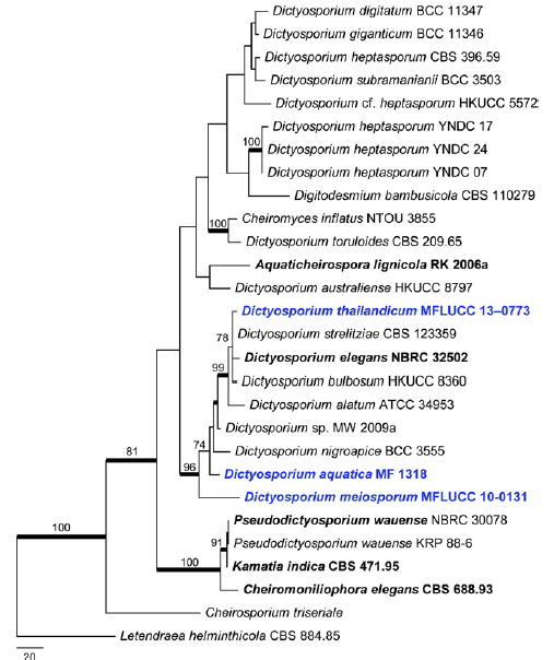
Fig. 1 Phylogram generated from Maximum Parsimony analysis based on combined ITS, SSU and LSU sequence data of cheirosporous and dictyosporious taxa. Parsimony bootstrap support values greater than 70 % are indicated above and below the nodes, and branches with Bayesian posterior probabilities greater than 0.95 are given in bold. The ex-types (reference strains) are in bold; the new isolates are in blue. The tree is rooted with Letendraea helminthicola CBS 884.85.
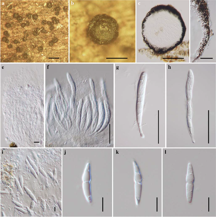
Fig. 2 Sexual morph of Dictyosporium meiosporum (holotype) a, b Ascomata occurring superficial on substrate c Section through of ascoma d Peridium e. Pseudoparaphyses f–h Asci i–l Ascospores. Scale bars: a=500μm, b, c=200μm, d=20μm, e, i-l=10μm, f-h=50μm.
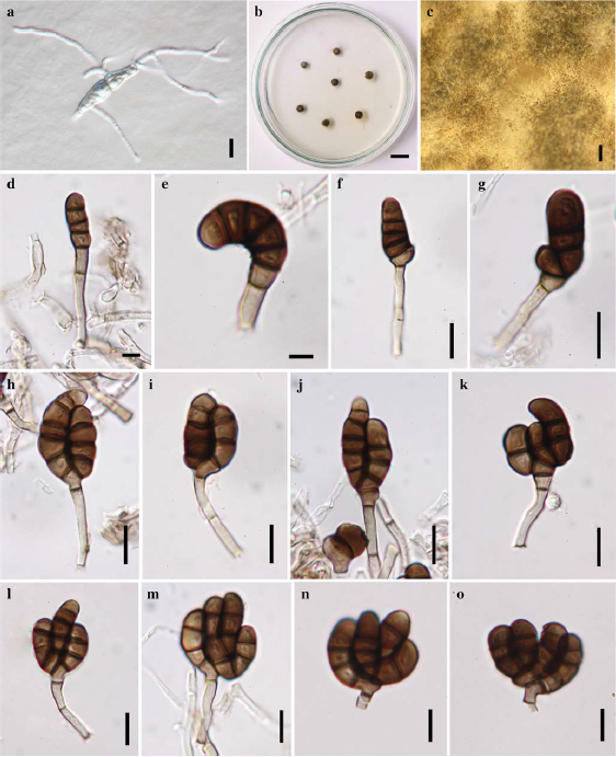
Fig. 3 Asexual morph of Dictyosporium meiosporum (ex-type culture) a Ascospore germination b Colonies culture on MEA c Vegetative hyphae d–i Conidiophores j–o Conidia with branches apical, dark brown, and several-septate. Note up to 4-conidium on conidiogenous locus. Scale bars: a, f, g-o=10μm, b=10 mm, c=500μm, d, e=5μm.