Dictyosporella guizhouensis J. Yang & K.D. Hyde, sp. nov.
Index Fungorum number: IF 556314; MycoBank number: MB 556314; Facesoffungi number: FoF 05999; Fig. 63
Etymology: Referring to the collecting location in Guizhou Province, China.
Holotype: MFLU 18-1505.
Saprobic on decaying twigs from freshwater habitats. Asexual morph: Colonies on natural substrate sporodochial, scattered, dark brown or black. Mycelium partly immersed, partly superficial, composed of septate, hyaline hyphae. Conidiophores macronematous, mononematous, compact, flexuous, simple or branched, mostly moniliform, with globose to subglobose, ellipsoid or clavate cells, hyaline, smooth-walled, up to 52 μm long. Conidiogenous cells monoblastic, integrated, terminal, globose, subglobose, ellipsoid or clavate, hyaline, smooth-walled, 9–17 × 6.5–12 μm ( x = 11.5 × 9.5 μm, n = 25). Conidia acrogenous, globose, subglobose, ellipsoid or irregular, muriform, with slightly irregular multi-euseptate, smooth, olivaceous to mid-brown, paler at the basal cell, 15–26 × 12.5–18.5 μm ( x = 19 × 16 μm, n = 50). Sexual morph: Undetermined. Material examined: CHINA, Guizhou Province, Anshun City, Gaodang village, 26°4.267′ N, 105°41.883′ E, on decaying wood submerged in the Suoluo river, 19 October 2016, J. Yang, GD 34–2 (MFLU 18-1505, holotype; HKAS 102160, isotype).
GenBank numbers: ITS: MK593606; LSU: MK593605; SSU: MK593611.
Notes: Dictyosporella, Junewangia and Sporidesmiella nested within Junewangiaceae in the phylogenetic tree based on a combined LSU and ITS sequence dataset (Fig. 64). However, Junewangia is shown to be polyphyletic which agrees with the result of Luo et al. (2019). D. guizhouensis clustered as a sister taxon to D. aquatica and D. thailandensis with full support (100% ML and 1.00 PP). D. hydei grouped with J. aquatica H.Y. Song & D.M. Hu, with full support (100% ML and 1.00 PP) and only one nucleotide difference, but only the LSU sequence data is available for D. hydei. Relationships between the genera and taxa in Junewangiaceae are awaiting to be resolved.
Morphological characters of the new taxon matched the generic concept of Dictyosporella. Septa in D. guizhouensis and D. hydei are slightly constricted while those in D. aquatica are strongly constricted. The conidial color is brown to black in D. aquatica, yellowish brown in D. hydei and olivaceous green in D. guizhouensis. In addition, D. guizhouensis has conspicuous hyaline globose to subglobose conidiogenous cells which are not observed in D. aquatica and D. hydei.
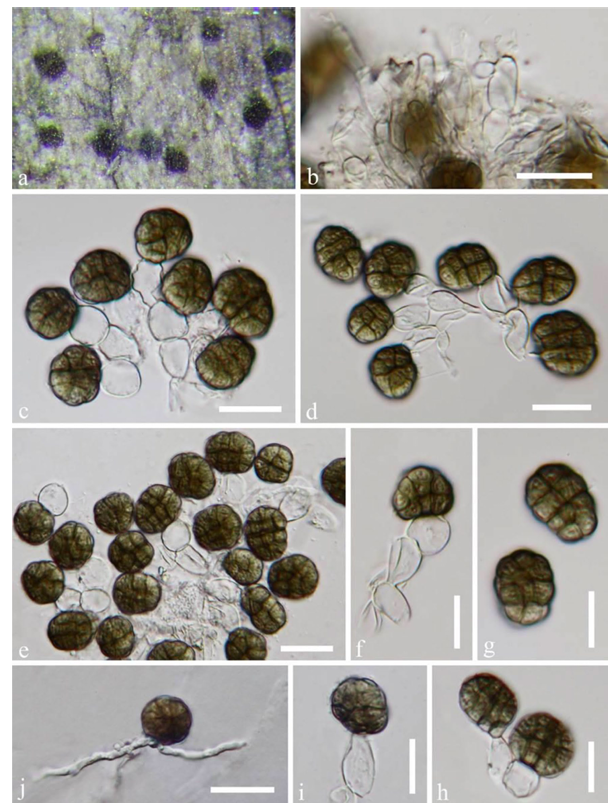
Fig. 63 Dictyosporella guizhouensis (MFLU 18-1505, holotype). a Colony on substrate. b Conidiophores. c–f Conidiogenous cells with conidia. g–i Conidia. j Germinated conidium on PDA. Scale bars: b–e, j = 20 μm, f–i = 10 μm
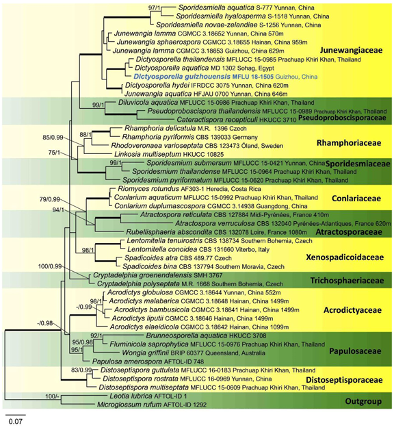
Fig. 64 Maximum likelihood tree of Diaporthomycetidae isolates based on a combined LSU and ITS sequence dataset. Bootstrap support values (ML) ≥ 75% and Bayesian posterior probabilities ≥ 0.95 are indicated above the nodes. Branches with 100% ML BP and 1.0 PP are shown as bold branches. The tree is rooted with Leotia lubrica (AFTOL-ID 1) and Microglossum rufum (AFTOL-ID 1292). The new species is in blue