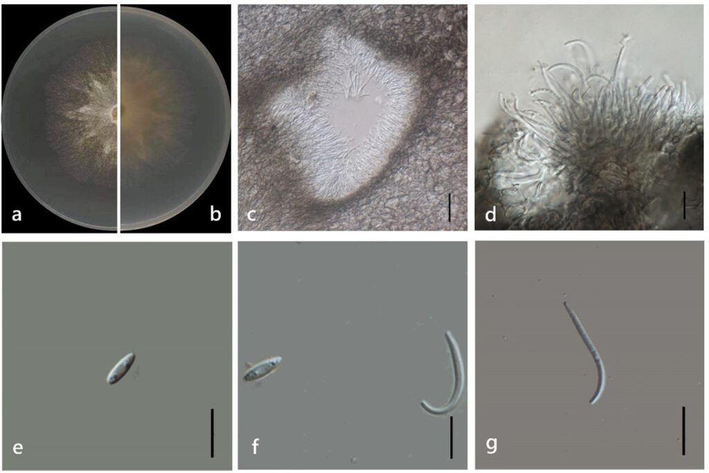
Diaporthe longiconidialis M. Luo, W. Guo, Manawas., M. P. Zhao, K. D. Hyde & C. P. You, sp. nov. (Figure 8)
MycoBank number: MB; Index Fungorum number: IF; Facesoffungi number: FoF 11352;
Etymology: In reference to its long spores.
Holotype: ZHKUCC 22-0040
Endophytic on Morinda officinalis root and stem. Sexual morph: not observed. Asexual morph: Pycnidia 40–400 × 30–160 μm (`x =150 ± 100 μm × 70 ± 40 μm), subglobose, flask or irregularly shaped, single or multiple cavities. Pycnidial wall consisting of sevearal layers of medium transparent textura globosa-angularis. Conidiophores hyaline, unbranched, densely aggregated, cylindrical-filiform, straight to sinuous, 1-septate. Conidiogenous cell phialidic, cylindrical, terminal and lateral, with slight taper towards apex. Alpha conidia 6–10 × 2–4 μm (`x = 8 ± 1 μm× 3 ± 0.3 μm), hyaline, fusiform, ellipsoid, long ellipsoid or lanceolate, contains at least one guttulate. Beta conidia 15–30 × 1–3 μm (`x = 20 ± 3 μm × 2 ± 0.3 μm), hyaline, linear, curved.
Culture characteristics: Colonies on PDA reach 85 mm diam. after 5 days. White cotton with some polygon mycelium as stars in the center, then with purple pigmentation. Fruiting body occurred from about 15 days, grey to dark black with orange surroundings. Reverse white with some purple pigmentation.
Material examined: China, Guangdong Province, Zhaoqing, isolated from a healthy root of Morinda officinalis. June 2020, W. Guo, dried culture (ZHKU 22-0040), and living culture (ZHKUCC 22-0058, ZHKUCC 22-0059–0066).
Habitat and host: Healthy stem and root of Morinda officinalis.
Known distribution: China (Zhaoqing, Guangdong Province).

Figure 8. Diaporthe longiconidialis (ZHKUCC 22-0058) (a) upper view of colonies on PDA; (b) Reverse view of colonies on PDA; (g) Horizontal section through a pycnidia; (h) Conidiogenous cells; (i,j) Alpha conidia; (j,k) Beta conidia. Scale bars: (g) =100 µm; (h–k) =10 µm.
