Cytospora calamicola Konta & K.D. Hyde. sp. nov.
MycoBank number: MB 559679; Index Fungorum number: IF 559679; Facesoffungi number: FoF 10825;
Etymology: Refer to both morphs were found on the host genus Calamus.
Holotype: MFLU 15-0262.
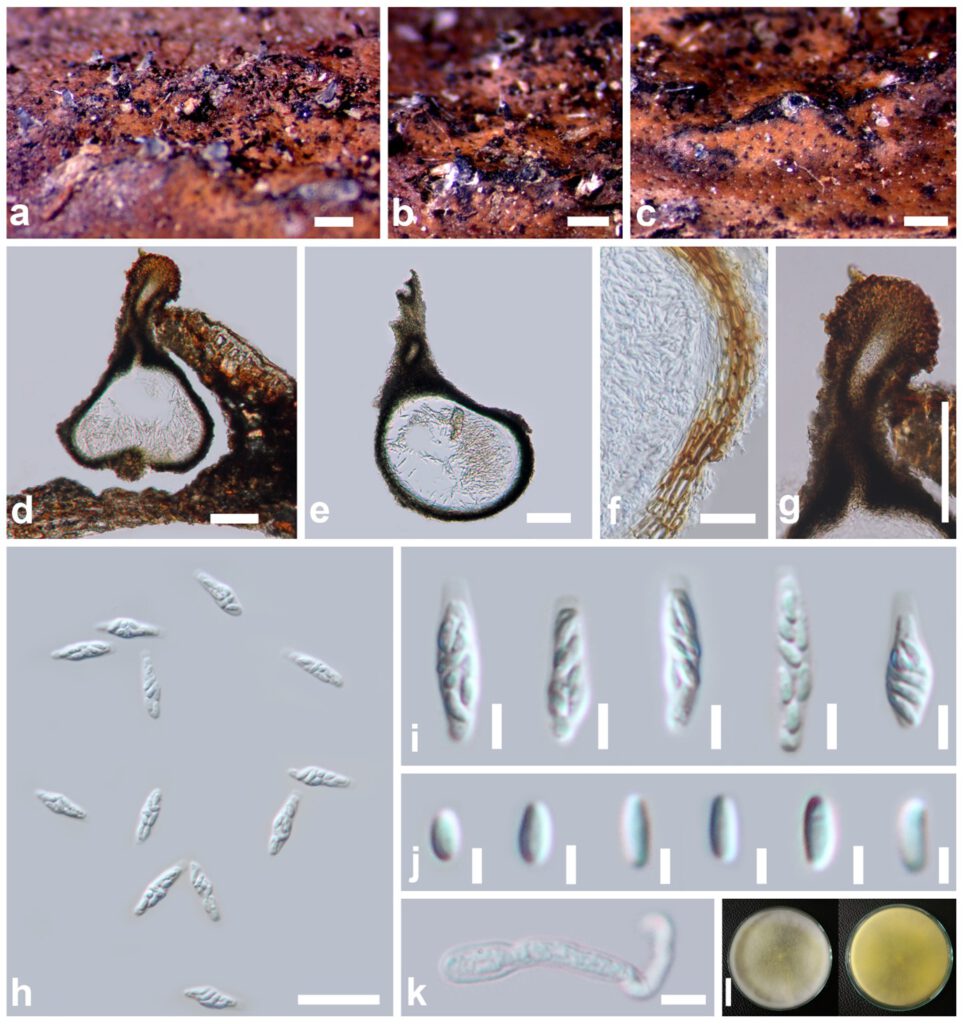
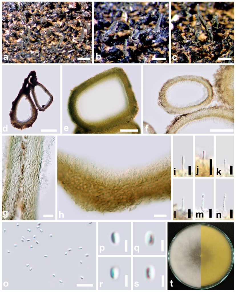
Saprobic on dead petiole of Calamus sp. (Arecaceae). Sexual morph: Ascomata 263–380 × 184–288 μm, scattered, immersed, eventually the neck erumpent, arising through cracks in bark epidermis, uniloculate, globose to subglobose or irregular, comprising whitish-yellow on host surface, long neck. Neck about 120–250 μm long, central, lined with periphyses. Peridium 13–16 μm wide, comprising an outer layer of yellow to pale-brown, thick-walled cells of textura prismatica and inner layer, hyaline, thin-walled layer of cells. Paraphyses absent. Asci 16–18 × 3.7–5.4 μm, (x̅ = 16.5 × 4.9 μm, n= 50), 8-spored, unitunicate, cylindrical to clavate, with thin-walled pedicel, without pedicellate, apex flat. Ascospores 3.3–5 × 1.1–2 μm, (x̅ = 4.3 × 1.7 μm, n= 100), biseriate or crowded, hyaline, fusiform or allantoid, oblong, unicellular, aseptate, smooth-walled. Asexual morph: Coelomycetous. Conidiomata 249–302 × 112–174 μm, appearing as brownish-yellow, pycnidia semi-immersed, solitary or aggregated, globose to subglobose, long neck. Peridium 10–13 μm wide, comprising pale-brown, thick-walled, cells of textura epidermoidea. Conidiophores erect, hyaline. Conidiogenous cells enteroblastic, proliferating percurrently to produce small, hyaline, ellipsoidal conidia. Conidia 1.7–2.8 × 1.1–1.7 μm, (x̅ = 1.8 × 1.3 μm, n= 20), oblong to fusiform, small, unicellular, hyaline and smooth-walled.

Fig XX Cytospora calamicola (MFLU 15-0259). a–c Appearance of ascomata on host substrate. d, e Section of ascoma. f Peridium. g Ostiolar neck. h, i Asci. j Ascospores. k Geminated ascospore. l Colony on MEA. Scale bars: a–c= 500 μm, d, e, g =100 μm, f = 20 μm, i = 5 μm, j, k = 2 μm.

Fig. XX Cytospora calamicola (MFLU 15-0262, holotype). a Appearance of conidiomata on host substrate. b, c Close up conidiomata. d–f Section of conidiomata. g Peridium. h–n Conidiogenous cells with conidia. o–s Conidia. t Colony on MEA. Scale bars: a = 1,000 μm, b, c = 500 μm, d = 100 μm, e, f = 50 μm, g–h = 10 μm, i–o = 5 μm, p–s = 2 μm.
