Conicomyces pseudotransvaalensis A. Hashim., G.= Sato & Kaz. Tanaka.
Index Fungorum number: IF551057, Facesoffungi number: FoF00486; Fig. 12
Etymology – named after its morphological similarity to Conicomyces transvaalensis R.C. Sinclair et al.
Holotype – HHUF 29956
Saprobic on Machilus japonica Siebold & Zucc. Sexual morph Undetermined. Asexual morph Conidiomata stromatic, synnematous, scattered, superficial, cone-shaped, black to dark brown, setose, up to 780μm high, 260–360μm wide at the base, cornuted with a head and a stipe; head slightly swollen, 65–100μm wide, bearing a concave conidial hymenium; stipe cylindrical or contorted, 30–100μm wide, composed of rectangular, thin-walled, brown, 1.5–2.5μm wide cells of textura porrecta. Setae arising from stroma or stipe, straight or curved, erect, septate, brown but pale at the apex, thick-walled, smooth, unbranched, up to 450μm long, acute and 2.5–4μm wide at the apex, 5–7μm wide at the base. Conidiophores arising from inner elements of the stipe, hyaline to pale brown, unbranched or branched, up to 40μm long. Conidiogenous cells phialidic, cylindrical, hyaline to pale brown, smooth, 16–88×2.5–3μm. Conidia 105–170×7.5– 10μm (x=150.8×8.8μm, n=50), L/W 12–22.7 (x=17.4μm, n=50), claviform, slightly obtuse at the apex, slightly truncate at the base, 15–22-septate, hyaline, smooth-walled, guttulate, bearing an unbranched appendage at the apex; appendage 40– 80μm long (x=59.8μm, n=50).
Culture characters – Conidia formed in culture are similar to those on natural substrate.
Material examined – JAPAN, Kagoshima, Yakushima, Yakusugi Land, dead twigs of Machilus japonica (Lauraceae), 15 March 2007, K. Tanaka & H. Yonezawa, GS 20 (HHUF 29956, holotype designated here); ex-type living culture, MAFF 244767.
GenBank accession numbers – ITS: LC001710; LSU: LC001708.
Notes – The genus Conicomyces was established to accommodate C. transvaalensis having synnematous conidiomata and apically appendaged conidia (Sinclair et al. 1983). Conicomyces currently contain three described species (Sinclair et al. 1983; Illman and White 1984; Seifert 1999), but no molecular studies have been undertaken for the genus. Morphologically C. pseudotransvaalensis is similar to C. transvaalensis in having large conidia more than 100μm long, but the latter has slightly long and slender conidia with more septation (122–200×5.5–7.5μm, L/W 25, 19–29-septate; Nag Raj 1993). Based on a megablast search, the closest hits to the 28S sequence of C. pseudotransvaalensis are Chaetosphaeria fuegiana (GenBank EF063574; Identities= 729/754 (96.7 %), Gaps 5/754 (0.7 %)), Chaetosphaeria hebetiseta (GenBank AF178549; Identities=723/754 (95.9 %), Gaps 5/754 (0.7 %)) and Chaetosphaeria dilabens (GenBank AF178557; Identities=720/751 (95.9 %), Gaps=5/ 751 (0.7 %)). These results clearly indicate that the genus is a member of Chaetosphaeriaceae (Sordariomycetes), as previously suggested by Hashimoto et al. (2015) based on morphological grounds. In Fig. 11, C. pseudotransvaalensis clusters in Chaetosphaeriaceae and is related to species of Chaetosphaeria sensu lato.
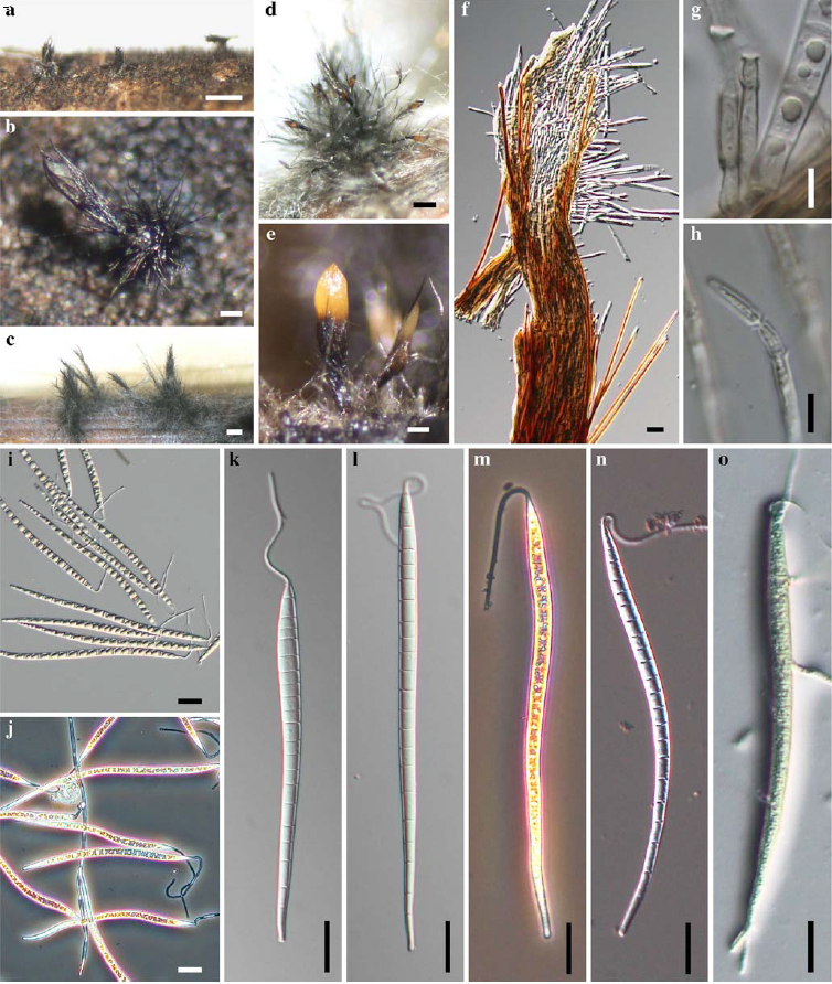
Fig. 1 Conicomyces pseudotransvaalensis (holotype). a, b Conidiomata on host surface c-e Conidiomata in culture f Conidioma in longitudinal section g, h Conidiogenous cells i–n Conidia o Germinating conidium a, b, f, h, k, l from HHUF 29956 (holotype); c-e, g, i, j, m–o from MAFF 244767 (ex-type isolate). Scale bars: a, c=500μm, b, e= 100μm, d=250μm, f, i–o=20μm, g, h=5μm.