Commelinaceomyces aneilematis (S. Ito) E. Tanaka, comb. nov. FIG. 1
MycoBank number: MB 831664;Index Fungorum number: IF 831664; Facesoffungi number: FoF;
Basionym: Ustilago aneilematis (as U. aneilemae) S. Ito, Trans Sapporo Nat Hist Soc 14:81. 1935.
Holotype: JAPAN. TOCHIGI: Kawachi-gun, Kaminokawa-machi, Isooka (present address), on Murdannia keisak (as Aneilema keisak), 11 Sep 1932, T. Watanabe & T. Yamada (SAPA2011).
Distribution: Japan, India, Taiwan
Sexual morph: Not observed.
Specimens examined: INDIA. MAHARASHTRA STATE: Kolhapur District, Gaganbawad, on M. keisak, 26 Oct 1986, S.R. Yadav (HUV 15783, HCIO 30106); loc. id., on M. keisak, Oct 1988, M.S. Patil (HUV 19045); Satara District, Kans, on M. keisak, 10 Oct 1989, M.S. Patil (HUV 19046). JAPAN. TOCHIGI: Kawachi-gun, Kaminokawa-machi, Isooka, on M. keisak, 11 Sep 1932, T. Watanabe & T. Yamada (isotype HUV 12340); ISHIKAWA: Hakusan, 36°30′36″N, 136°35′16″E, on M. keisak, 5 Oct 2015, E. Tanaka (TNS-F-87155, culture MAFF 246963); Nonoichi, 36°30′39″N, 136°36′06″E, on M. keisak, 9 Oct 2015, E. Tanaka (TNS-F-87156, culture MAFF 246964); Kanazawa, 36°37′48″N, 136°40′20″E, on M. keisak, 10 Oct 2015, E. Tanaka (TNS-F-87157, culture MAFF 246965). TAIWAN. HSINCHU: Near Hsinchu, road to Wu Zhi Shan, on M. keisak, 28 Mar 1999, C.-J. Chen, R. Kirschner & M. Piepenbring (HUV 21510).
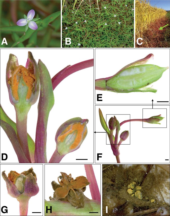
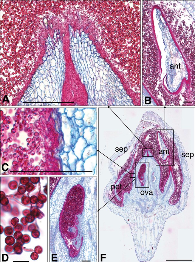
Notes: Commelinaceomyces aneilematis forms sori in the flowers of M. keisak, which is a native plant species of eastern Asia, where it is found in damps soils and wet areas (FIG. 1). Infection by C. aneilematis appeared systemic, as most flowers on stems of infected plants formed sori. Infected flower buds were enlarged and filled with bright yellow to orange conidial masses (FIG. 1D) that ruptured to expose orange to olivaceous green conidial masses (FIG. 1G and H). The fallen spore mass floated on the surface of the water and dispersed (Fig. 1I). Stamens were not affected, and neither hyphae nor conidia were found in the anthers (FIGS. 1H and 2). Sections of infected flowers stained with PAS showed that conidial masses filled the space between the floral parts (FIG. 2). Ovaries were covered with mycelia, and sometimes hyphae and conidia were observed in the ovaries (FIG. 2F). It appeared that hyphae invaded from the stigma (FIG. 2A).
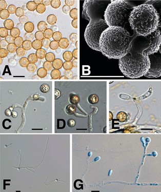
The conidia of C. aneilematis are globose to sub- globose, 9–11.5 µm diam, with a densely verruculose wall 0.7–1.2 µm wide (FIG. 3A, B). Conidia form at the apex of conidiogenous cells, in contrast to other members of the Ustilaginoideae, which form conidia pleurogenously. The conidiophores of C. aneilematis terminate in dichotomous or trichotomous branches (FIG. 3C–E). Conidiogenous cells are cylindrical or clavate. The conidia germinate readily on water agar (FIG. 3F). On PDA, mycelial growth is sympodial, with spherical, ovoid, and hyaline secondary conidia formed at the apex of simple conidiogenous cells (FIG. 3G). The morphological characteristics of the specimens of C. aneilematis matched the description of U. aneilematis from Japan (SUPPLEMENTARY TABLE 2). The DNA sequence analyses revealed that C. aneilematis belonged to the clavicipitaceous tribe Ustilaginoideae. There was one nucleotide insertion or deletion in the ITS1 sequences of three isolates collected from Japan.

Figure 1. Commelinaceomyces aneilematis and its host Murdannia keisak. A. Flower of M. keisak. Host plants were found in rice paddy fields in late summer with 3-petaled, pink or purple-white flowers at the shoot apices or leaf axils. B. Dense cover of plants of M.keisak in a paddy field. C. Mature leaves and shoots of M. keisak turn red or purple and cover rice plants (arrow). D–F. Infected shoot of M. keisak (TNS-F-87155). Most flowers on an infected shoot are enlarged and filled with bright yellow to orange-yellow, dusty spore masses. D. Cross-sections of immature infected flowers filled with conidia. E. Cross-section of a healthy ovary. G and H. Matured sori with emergent orange to olive-green spore masses. I. Spores floating on water. Bars: 1 mm.

Figure 2. Sections of a flower of Murdannia keisak infected with Commelinaceomyces aneilematis stained with Alcian blue and periodic acid– Schiff (PAS). Magenta color indicates fungal materials. A. Hyphae in the gap at the apex of the pistil. B. Anther was not infected by fungi. C. Surface of the ovary covered with mycelia. D. Thick-walled conidia formed on conidiogenous cells. E. Hyphae and spores in an ovary. F. Spore mass in spaces between floral parts. ant = anther; ova = ovary; sep = sepal; pet = petal. Bars: A, B, C, E = 100 µm; D = 10 µm; F = 1 mm

Figure 3. Conidia of Commelinaceomyces aneilematis. A. Thick- walled conidia under light microscopy. B. Thick-walled conidia under scanning electron microscopy. C–E. Thick-walled conidia and conidiogenous cells. C. Mature conidiogenous cells with developing conidia. D. Matured thick-walled conidia at the apex of conidiogenous cells. F. Germinated thick-walled conidia on water agar. G. Secondary conidia with conidiophores on PDA stained with lactophenol cotton blue. Bars = 10 µm.
