Colletotrichum fructicola Prihast., L. Cai & K.D. Hyde, in Prihastuti, Cai, Chen, McKenzie & Hyde, Fungal Diversity 39: 96 (2009).
MycoBank number: MB 515409; Index Fungorum number: IF 515409; Facesoffungi number: FoF 06767;
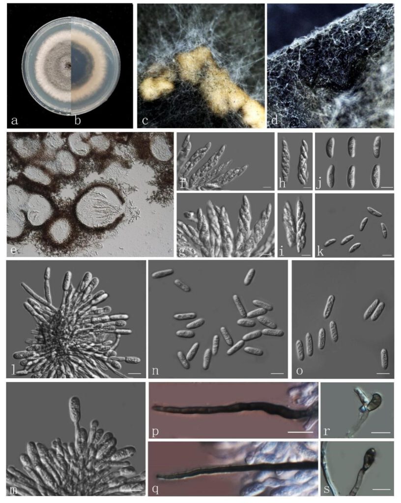
Micromorphology: Associated with leaf spot of Zamia furfuracea. Sexual morph: ascomata greyish black, globose to subglobose, with hairs, semi-immersed or completely immersed in PDA. ascoma (45–)50–65 (–80) × 10–12 μm (ave. 62 × 10 μm, n=30) long, thin-walled,6-8 spored, clavate or cymbiform. Ascospores (13–) 15–18 (–20) × 5–6 (–7) (ave. 16 × 6μm, n=50) long, one-cells, large guttulate at the centre, slightly curved to curved with rounded ends. Asexual morph: Setaria in acervuli on filter paper, abundant, long, dark brown, smooth-walled, with two or more septa, base truncate, tip sharp, (44–) 56 × 2–4 μm (ave. 60 × 3 μm, n=7). Conidiophores hyaline, smooth-walled to verruculose, aseptate, unbranched. Conidia are formed from the tip of the conidiophore, one-celled, cylindrical with rounded ends, some acervulied ends, contents appearing, granular, 16–19 × (4–) 5–6 μm (ave. 17 × 5 μm, n=50). Appressoria in slide cultures, formed from branched mycelia, terminal, brown to dark brown, near rhombus to irregular shape, (8–) 10 × (5–) 6–7 μm (ave.10 × 6 μm, n=10).
Culture characteristics: Colonies on PDA, reaching 65mm diam. after 7 days at 25 ℃, growth rate per day 5 mm/d (n=4), grey to green, the edge is white, regular, reverse dark green, the edge is white with mycelia flourishes, felt-like, with orange conidial masses.
Material examined: China, Guangdong province, Shenzhen Botanical Garden, on living Zamia furfuracea L.f. ex Aiton, 30th November 2020, J.W. Chen, Z.H. Zhang and C. Chen (new host record) (dried cultures ZHKU 21-0074 and ZHJU 21-0075) and living cultures ZHKUCC 21-0081 and ZHJUCC 21-0082.
Notes: our species is introduced based on multigene analysis of GAPDH, ACT, CHS-1 and TUB2 sequence data. The species is phylogenetically closely related to Colletotrichum fructicola with moderate support (87% in ML, 97% in MP) (Fig. 42). Compared the morphology of our isolates with C. fructicola described by Prihastuti et al. (2009), they are similar in the sexual morph. The difference is the size of conidia, our isolates are 16–19 × (4–) 5–6 μm (x̅ 17 × 5 μm, n=50), which is longer and wider than the later, 9-14 × 4.5 um (x̅ 12 × 4 n = 180) (Prihastuti et al, 2009).

Fig. Colletotrichum fructicola (ZHKUCC 21-0081: New host record on Zamia furfuracea) a, b Upper and reverse view on PDA after seven days. c Spore masses. d, e Ascomata. f–i Asci. j, k Ascospores. l, m Conidiomata and conidiogenus cells. n,o conidia. p, q Setae. r, s appressoria. Scale bars: f–s = 10 µm.
