Chaetosphaeria rivularia Réblová & J. Fourn. [as ‘rivularia‘], in Ariyawansa et al., Fungal Diversity: 10.1007/s13225-015-0346-5, [119] (2015)
Index Fungorum number: IF551489; Facesoffungi number: FoF00975;
Etymology − Rivularis (L), referring to rivulets, the natural habitat of the fungus.
Holotype − PRM 933847
Saprobic on decaying wood submerged in freshwater. Sexual morph: Ascomata non-stromatic, semi-immersed, scattered to gregarious, often with confluent walls, venter 200–250 μm diam., 250–330 μm high, subglobose to broadly conical, brown, glabrous, papillate, opening by a rounded pore. Ostiole central, periphysate. Peridium leathery to fragile, carbonaceous, two layered, 30–60 μm thick near apex and at the sides, up to 50–60 μm thick at the base; outer wall composed of brown, brick-like cells of textura prismatica, cells opaque in the upper part, pale brown at the base, towards the interior grading into several rows of thin-walled, hyaline, flattened cells. Hamathecium composed of abundant, persistent, septate, hyaline paraphyses ca. 3.5–4.5 μm wide, tapering to 2.5−3 μm, longer than the asci. Asci (72–)75–95(−98)×7– 8.5(−9) μm (x ± SD=86.7±8.2×8±0.6 μm), 8-spored, unitunicate, cylindrical-clavate, rounded, slightly tapering at the apex, short-pedicellate, with a distinct, non-amyloid apical annulus, ca. 1–1.3 μm high, 2–2.5 μm wide. Ascospores (15–)16−19×3–3.5 μm (x ± SD=16.8±1.3× 3.4±0.3 μm), ellipsoid to narrowly fusiform, slightly curved, 1-septate, septum indistinct, not constricted at the septum, hyaline to yellowish grey, smooth-walled, arranged obliquely uniseriate, often two-seriate only in the upper sporiferous part, with a hardly defined mucilaginous sheath swelling and diffusing in water. Asexual morph: Thozetella-like formed only in the axenic culture.
Culture characters − Colonies slow growing, reaching 10– 15 mm diam. on PCA after 14 days at 25 °C. Colony circular, felty, zonate, brown-grey in the centre, brown towards the margin, reverse dark brown. Vegetative hyphae 2–3.5 μm wide, smooth, subhyaline or pale brown, thin-walled, septate, sparsely branched. Sporulation appears in a month. Synnemata scattered in circular zones around the centre and at the margin of the colony, composed of dark brown stalk bearing a slimy, glistening, white-grey mass of conidia. Stalk ca. 10–30 μm wide, composed of a brown, septate, sparsely branched conidiophores densely bunched and partly fused, ending in a monophialide, topped by a subglobose mass of conidia and sterile awn-shaped cells, so called microawns. Microawns produced from conidiophores and liberated with the conidia, straight to sigmoid or irregularly curved, basal part thin-walled, tapering towards the tip, thick-walled, pointed at the tip, 0–3-septate, hyaline, 65–130 μm long, 4–6.5 μm wide at the broadest point near the base, rigid, refractive. Conidiophores macronematous, up to 100 μm long, 2.5– 3.5 μm wide, pale brown, septate, branched at intervals. Conidiogenous cells phialides, 16–35×2–2.4 μm (x ± SD= 23.8±7.5×4.5±0.4 μm), integrated, terminal, cylindrical to clavate, tapering to ca. 1.5 μm, bearing a single conidiogenous locus in a minute, indistinct collarette. Conidia (9.5−)11−13× 1.8−2.5 μm (x ± SD=12.2±0.7×2.2±0.2 μm), lunate to suballantoid, hyaline, aseptate, at each end slightly narrowed and provided with a single filiform setula 2.8−4.5(−5.5) μm long.
Material examined − FRANCE, Midi-Pyrénées, Ariège, Rimont, Grand Bois forest, Maury brook, 650 m a.s.l., submerged wood of Fagus sylvatica associated with Minutisphaera japonica, 17 November 2009, J. Fournier J.F. 09308 (PRM 933847, holotype); ex-type living culture CBS 127686; Ibid., 24 November 2009, J. Fournier J.F. 09316 (PRM 933847). Ibid., 8 December 2009, J. Fournier J.F. 09327.
Notes − Chaetosphaeria rivularia was repeatedly collected on wood submerged in freshwater in southern France. Noconidiophores were observed on the host. The living culture derived from ascospore isolates from a fresh collection of C. rivularia yielded synnematous conidiophores with long, thick-walled and pointed microawns and setulose, aseptate Thozetella-like conidia. The relationship between Chaetosphaeria Tul. & C. Tul. and the synnematous dematiaceous hyphomycete Thozetella Kuntze was suggested by Paulus et al. (2004), based on the phylogenetic analysis of ITS rDNA sequence data. Thozetella comprises to date 19 species occurring primarily as saprobes on decaying wood, herbs, litter, palm fronds and rarely as endophytes. The only species so far reported from wood submerged in freshwater is T. nivea (Berk.) Kuntze, the type species (Sivichai et al. 2002). In our phylogenetic analysis based on ITS and nuc28S rDNA
sequences of 82 members of the Chaetosphaeriales a close relationship of C. rivularia with other permanently asexual Thozetella species is well-supported.
Chaetosphaeria rivularia resembles C. ciliata, C. ovoidea and C. tortuosa (Holubová-Jechová 1973; Réblová et al. 2006; Réblová and Seifert 2008) in the size and shape of the ellipsoid to narrowly fusiform, often curved, septate ascospores and setulose conidia. The three latter species differ from C. rivularia by 3-septate ascospores and formation of the Menispora asexual morphs with mononematous conidiophores. The ascospores of C. rivularia are 1-septate, the middle
septum is rather indistinct. Ascospores possess a mucilaginous sheath that is visible only in fresh material; the sheath diffuses in water and it is visible when observed in India ink. The asexual morph of C. rivularia is characterized by synnematous branching conidiophores, long, pointed, curved to sigmoid microawns that contain 1–3 septa, but sometimes appear as septate and aseptate conidia. It resembles T. gigantea in the morphology of microawns and conidia, but the latter species differs by longer conidia and longer setulae (Paulus et al. 2004).
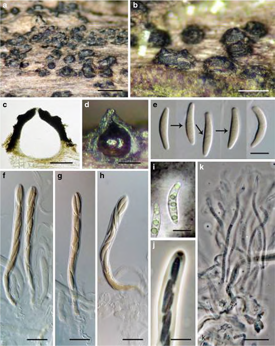
Fig. 1 Chaetosphaeria rivularia (holotype) a, b Ascomata c, d Vertical section of ascoma e, i Ascospores with indistinct middle septum (arrows indicate septum), e in Melzer’s reagent, i in India ink) f–h Asci j Ascal apex with an apical annulus k Paraphyses. Scale bars: a=500 μm, b= 250 μm, c, d=100 μm, e, i, j=10 μm, f–h, k=25 μm