Ceratosphaeria yunnanensis C.F. Liao & Doilom sp. nov. Fig. ###
MycoBank number: MB 559274; Index Fungorum number: IF 559274; Facesoffungi number: FoF 10583;
Etymology: ‘yunnanensis’ referring to the species was collected from Yunnan province, China.
Holotype: KUMCC 21-0013 (112878)
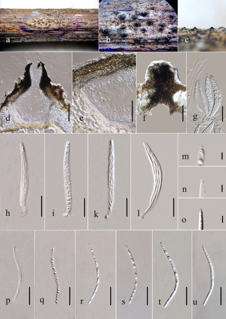
Saprobic on stem of Heteropanax fragrans. Sexual morph: Ascomata 111 – 215 × 130 – 244 μm ( = 164 × 196 μm, n = 20), solitary, deeply immerse, neck erumpent through the host surface, globose to subglobose, brown to yellowish. Neck 60 – 98 μm wide ( = 82 μm, n = 20), from the bottom to the apical part, the color changes to brown or black to yellowish brown, surface smooth. Peridium 20 – 52 μm wide ( = 41 μm, n = 30), composed of 3-6 layers of brown cell to yellowish brown cell of textura prismatica, thick-walled. Paraphyses 2.3 – 10.5 μm wide ( = 6.5 μm, n = 30), hyaline, unbranched, septate, border at the base, tapering to the end, longer than asci. Asci 84 – 123 × 4 – 13 μm ( = 106 × 8 μm, n = 30), 8-spored, unitunicate, faciculate, cylindrical, narrow, thin walled with an apical ring. Ascospores 77.8 – 109 × 1.3 – 2.8 μm ( = 88 × 2 μm, n = 30), straightly or curved, C-shaped or S-shaped, filiform, aseptate, hyaline, tapering at both ends, with minute guttules in each cell, becoming smooth at maturity. Asexual morph: undeerminted.
Culture characters: Ascospores germinating on PDA within 24 hours. Colonies on PDA reaching 1.2 – 2.1 cm diameter after 10 days at 25 ± 2 °C, umber to pale umber at the edge, white surface forms a circular ring in the middle. Colonies rough, mycelium dense. Reverse red to yellow, no pigment produce.
Material examined: CHINA, Yunnan Province, Kunming, on dead stem of Heteropanox fregans, 12 August 2020, C.F. Liao (KUN–HKAS 112878, holotype), ex-type culture KUMCC 21-00.
GenBank number: SSU: ######, LSU: ######, ITS: ######, TEF: ######.
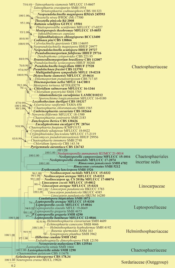
Notes: In phylogenetic analysis, our taxa are closely to Ceratosphaeria lignicola (Fig##). In morphology, Ceratosphaeria yunnanensis is similar to species of Ceratosphaeria lignicola with respect to the deeply immerse ascomata in the substate. However, Ceratosphaeria yunnanensis differs from C. lignicola by its smaller ascomata (111 – 215 × 130 – 244 μm vs 390–470 × 500–600 μm) and thick perdium (20 – 52 μm vs 13.5–17.5 μm). Ceratosphaeria yunnanensis comprised with Ceratosphaeria lignicola by sequence of LSU and TEF revealed 10 and 23 different nucleotides (totally have 750 and 846 nucleotides). Hence, we introduced Ceratosphaeria yunnanensis to a new taxa based on available morphology and phylogenetic tree.

Fig. ### Phylogenetic tree derived from maximum likelihood analysis of a combined ITS, LSU of 80 sequences and the aligned dataset was comprised of 1520 characters including gaps (ITS: 1-545 and LSU: 546-1520). A best scoring RAxML tree was established with a final ML optimization likelihood value of -20765.086284. The matrix had 900 distinct alignment patterns with 30.81% undetermined characters or gaps. Estimated base frequencies were found to be: A = 0.221860, C = 0.274373, G = 0.319603, T = 0.184164; substitution rates AC = 1.570428, AG = 2.097555, AT = 1.612952, CG = 1.187456, CT = 7.503192, GT = 1.000000. Gelasinospora tetrasperma (CBS 178.33)and Neurospora crassa (MUCL 19026) were used as outgroup. Numbers above branches are the bootstrap statistics percentages (left) and Bayesian posterior probabilities (right). Branches with bootstrap values Bootstrap support values for ML and MP ≥75% and ≥ 0.95 are shown at each branch and the bar represents 0.06 substitutions per nucleotide position. Hyphen (-) represents support values ≤ 75%/0.95. Ex-type strains are in black bold, newly generated sequence is indicated in blue and bold for new taxa.

Fig. ### Pseudocapulatispora fragrantis (KUN–HKAS 107635, holotype). (a–c). Appearance of ascomata on substrate. (d). Section through ascoma. (e). Peridium. (f). Ostiole’ (g). Paraphyses with asci. (h–l) Asci. (m–o) polar mucilaginous appendage. (p–u). Ascospores. Scale bars: (d) = 500 μm, e = 200 μm, (f, g–l, p–u) = 100 μm, (m–o) = 10 μm.
