Caryospora aquatica Huang Zhang, K.D. Hyde & Ariyawansa.
Index Fungorum number: IF551418; Facesoffungi number: FoF00958; Fig. 2
Etymology– in reference to the aquatic habitat.
Holotype – MFLU 11-1083
Saprobic on submerged wood. Sexual morph: Ascomata 350 – 600 μm high, 450 – 750 μm diam., solitary or clustered, erumpent, nearly superficial, hemisphaerical, base flattened, dark brown to black, carbonaceous, with ostiolate papilla. Papillae 50 μm long, 150 μm diam., apex truncate. Ostiole central, relatively broad, up to 0.1 mm diam., circular, brown to black. Peridium up to 90 μm wide at the sides and 10 μm wide at the base, strongly carbonized, composed of a black amorphous layer that cannot be differentiated, whose cells are rectangular, uninucleate, and often occluded. Hamathecium comprising numerous, up to 1 μm wide, filiform, trabeculate, anastomosing pseudoparaphyses, embedded in a gelatinous matrix. Asci 160 – 190 × 60 – 80 μm (x̄ = 179.6 × 63.4 μm, n = 12), 8 – spored, bitunicate, fissitunicate, broadly cylindric – clavate, usually slightly curved, pedicellate, apically rounded, with an ocular chamber. Ascospores 44 – 52 × 20 – 27 μm (x̄ = 47.4 × 23.4 μm, n = 15), 2 − 3 – seriate, 1 – septate, broad – fusiform and hyaline when young, becoming irregularly diamond shaped and dark brown at maturity, ends acute, slightly constricted at the septum, with polar germ pores at each end, relatively thick – walled, smooth – walled, with large globule in each cell, surrounded by narrow thin gelatinous mucilaginous sheath. Asexual morph: Undetermined.
Material examined – THAILAND, Chiang Rai Province, Hui Kang Pla Waterfall, on submerged wood, 18 January 2010, Huang Zhang (MFLU 11-1083, holotype); ex-type culture, MFLUCC 11-0008.
Notes – Caryospora minima Jeffers was first mentioned by Ellis and Everhart (1892) from peach stones, but they thought it was an 8-spored version of the type species of Caryospora, C. putaminum. Jeffers (1940) redescribed it as a new species when studying C. putaminum. The perithecia in C. minima are smaller than in C. putaminum (400 – 750 vs. 500 – 1200 μm). In this paper, we introduce a new species C. aquatica from submerged wood in freshwater. Caryospora aquatica is similar to the type species, C. putaminum in having erumpent, superficial, dark brown to black, carbonaceous, ostiolate ascomata, a thick and carbonized peridium, and relatively large and thickwalled ascospores, but can be distinguished in the smaller ascoma (350 – 600 vs. 500 – 1200 μm in C. putaminum) and 8 – spored asci (2 – spored in C. putaminum). Caryospora aquatica also differs from C. minima in its freshwater habitat. At maturity, the ascospores of C. minima are light brown with 3 septa, while in C. aquatica they become irregularly diamond-shaped and dark brown with polar germ pores. Caryospora easily produces perithecia in culture. Jeffers obtained perithecia in C. minima in peach-stone culture and we also found these in C. aquatica on PDA media. Under moist condition the spores easily discharge through the circular ostiole and form the plume on top of the perithecium.
In the phylogenetic tree (Fig. 1), C. aquatica clusters with a putatively named strain of C. minima, which was also isolated from submerged wood, and is sister to Acrocordiopsis clade. Caryospora has been collected from wood in freshwater on numerous occasions (Cai et al. 2003; Hyde et al. 1998; Kurniawati et al. 2010; Luo et al. 2004; Zhang et al. 2011) and was usually named as C. minima. The Caryospora species from freshwater are likely to comprise several taxa as has been shown for the freshwater species of Helicascus (Zhang et al. 2013a, b, 2014; Shearer 1993; Cai et al. 2003). This is the first time to confirm the natural placement of Caryospora species with verified strains, at the family level with molecular data.
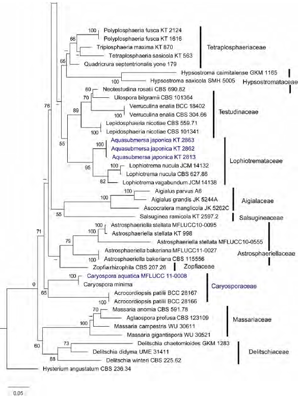
Fig. 1 (continued)
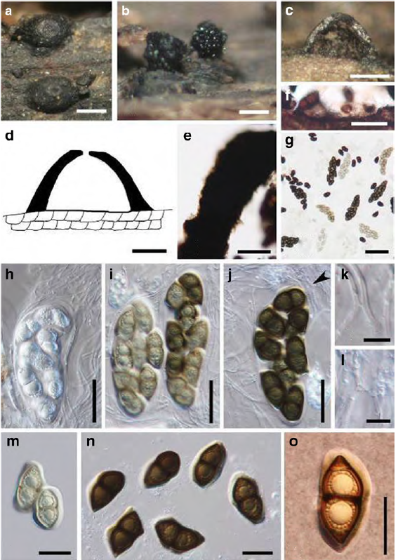
Fig. 2 Caryospora aquatica (holotype) a, b Appearance of ascomata on the host surface. Note the plume of ascospores in b. c Section of an ascoma on wood d Section of ascoma e Peridium f Base of ascoma g Different ages of asci h Young ascus with trabeculate pseudoparaphyses i Nearly mature ascus j Mature ascus with anastomosing pseudoparaphyses (arrowed) k Trabeculate pseudoparaphyses l Anastomosing pseudoparaphyses m, n, o Ascospores. Note the polar germ pores in o. Scale bars: a, b, c, g = 200 μm, d, f = 100 μm, e, h, i, j = 50 μm, k, l = 10 μm, m, n, o=30 μm.