Camarosporium clematidis Wijayawardene, Tangthirasunun, Camporesi & K.D. Hyde, in Wijayawardene, Bhat, Hyde, Camporesi, Wikee, Ariyawansa, Chethana, Tangthirasunun, Chukeatirote & Wang, Phytotaxa 183(1): 19 (2014)
Index Fungorum Number: IF 807934, MycoBank Number: MB 807934, Facesoffungi number: FoF 00001
Etymology — Named after the host genus on which the fungus occurs.
Saprobic on dead and hanging branch of Clematis vitalba. Sexual state: Not observed. Asexual state: Conidiomata 370–425 μm diam., 350–380 μm high, pycnidial, immersed in the host, dark brown to black, unilocular, ostiolate, separate or aggregated. Conidiomata wall 5–10 layered, 45–95 µm thick, cells thick-walled, dark brown, composed of cells of textura angularis. Ostiole single, central, circular, papillate. Conidiophores absent. Conidiogenous cells enteroblastic, annellidic, smooth, hyaline, formed from the innermost layer of pycnidial wall cells. Conidia 10–17 × 7–9 μm (mean 13.6 × 8.1 μm, n = 20), oblong, muriform, with 2–3 transverse and 1 longitudinal septa, slightly constricted at septa, rounded at both ends, smooth, variable in colour, medium to dark brown.
Colony characteristics — On PDA white to very light brown from above and reverse, with sparse mycelium, flat, rhizoidal at the margin, attaining a diam. of 3.5 cm in 7 days at 27 ºC.
Material examined — ITALY. San Martino-Predappio: Forlì-Cesena, dead and hanging branch of Clematis vitalba, 25 March 2012, Erio Camporesi (MFLU13–0336, holotype); ex-type living culture = MFLUCC 12–0354 = ICMP 20051 = HGUP N_1.
Notes — Sutton (1980) and Ellis & Ellis (1985) listed coelomycetes that inhabit Clematis, viz. Asteromellopsis insculpta H.E. Hess & E. Müll., Ceratopycnis clematidis Höhn., Hoehneliella perplexa Bres. & Sacc., Phoma lingam (Tode) Desm., Phomopsis demissa (Sacc.) Traverso and Septoria clematidis Roberge ex Desm. Among these taxa, Ceratopycnis clematidis (from Clematis vitalba) is the closest taxon to our collection. Both species have enteroblastic conidiogenesis and brown conidia. However, Ceratopycnis clematidis has only transverse eusepta (phragmospore) (Sutton 1980) while our collection has conidia with transverse and longitudinal septa (muriform conidia). In molecular data analyses, our strain groups with Camarosporium quaternatum, the type species of Camarosporium. There appears to be no Camarosporium spp. described from Clematis vitalba, hence, we introduce our collection as a new species.
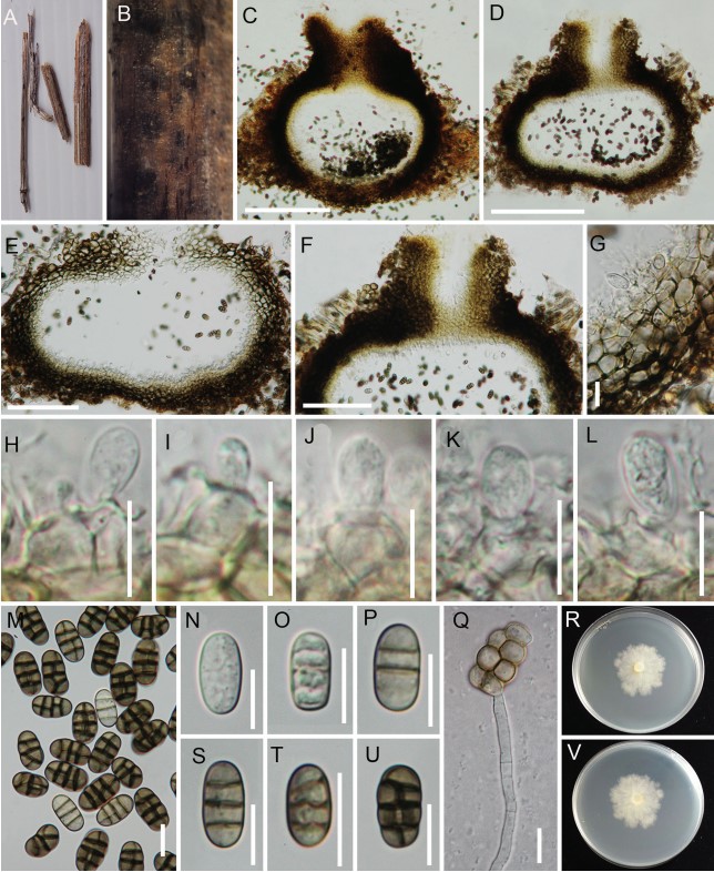
Figure 2. Camarosporium clematidis (MFLU13–0336). A. Conidiomata on dead branch of Clematis vitalba. B. Conidiomata on host surface. C, D. Longitudinal section of a conidioma with ostiole. E, G. Longitudinal section of a conidioma wall. F. Longitudinal section of ostiole. H–L. Conidiogenous cells with developing conidia; note annellations in J and L. M–S. Conidia. T. Germinating conidium. U–V. Colonies on PDA; U. From top; V. From reverse. Scale bar: C, D = 200 μm, E, F = 100 μm, G–T = 10 μm