Brachiampulla verticillata (B. Sutton & Hodges) Réblová & Hern.-Restr., in Réblová, Kolařík, Nekvindová, Miller & Hernández-Restrepo, Microorganisms 9(4, no. 706): 45 (2021)
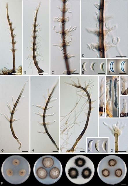
Index Fungorum number: IF 837809; MycoBank number: MB 837809; Facesoffungi number: FoF 15024 (Figure 19).
Basionym – Selenosporella verticillata B. Sutton & Hodges, Nova Hedwigia 29: 602.1978 (1977).
Synonym – Zanclospora urewerae J.A. Cooper [as “ureweri”], N. Z. J. Bot. 43: 344. 2005.
Description on the natural substrate: Colonies effuse, hairy, dark brown, whitish-brown when sporulating. Asexual morph: Conidiophores 120−173 × 4−5.5(–6) µm, erect, single or in fascicles, cylindrical to subulate, base bulbose, setiform, straight or gently bent, unbranched, septate, dark brown towards the base, paler towards the apex, smooth, apical cell subhyaline to light brown, often fertile. Conidiogenous cells polyphialidic, 11−16 × 4−6 µm, venter 6–8 µm long, the upper part above venter sympodially extending 4.5−9.5 × 1.5−2 µm, with numerous lateral openings with minute collarettes, indeterminate, ampulliform to lageniform, discrete, lateral, arranged singly or in groups of 2−5 in whorls below the transverse septa, or integrated, disposed of terminally, light brown towards the base, subhyaline towards the apex, smooth, diverging from the conidiophore. Conidia 6.5−8 × 1.5−2 µm (mean SD = 7.2 ± 0.4 × 1.8 ± 0.2 µm), lunate, hyaline, tapering at both ends, acute apically, with a basal scar, smooth, aggregated in white slimy heads. Sexual morph: Not observed.
Culture characteristics – On CMD colonies 14–15 mm diam, circular, flat, margin finely fimbriate, cobwebby to sparsely floccose becoming mucoid towards the margin, amber-beige, with a light beige outer zone of submerged growth, reverse light brown. On MLA colonies 24–25 mm diam, circular, convex centrally, flat margin, margin finely fimbriate, lanose, floccose, furrowed at the centre, zonate, beige with a beige-brown intermediate zone, dark brown to russet at the margin, reverse dark brown. On OA colonies 23–25 mm diam, circular, flat, margin subsurface, lobate, lanose, floccose becoming mucoid towards the margin, beige-brown with a black outer zone of submerged growth, reverse black. On PCA colonies 15–17 mm diam, circular, slightly raised, margin entire, lanose, floccose becoming cobwebby to locally mucoid at the margin, beige, dark brown at the margin with a russet outer zone of submerged growth, reverse dark brown. Sporulation was abundant on CMD, OA (restricted to the inoculation block and the centre of the colony), moderate on PCA, and absent on MLA.
Description on PCA − Colonies effuse, hairy, vegetative hyphae subhyaline to brown, septate, branched, 1.5–3 µm wide. Asexual morph: Conidiophores, conidiogenous cells and conidia as on the natural substrate. Conidiophores 105−168 × 3.5−4.5 µm, apical cell fertile, terminated into a phialide or a whorl of several phialides. Conidiogenous cells polyphia- lidic, 13.5−25 × 4−6 µm, venter 6–8 µm long, the upper part above venter sympodially extending (6−)7−17.5 × 1.5−2.5 µm, occasionally with 1–2(–3) percurrent elongations, discrete, lateral, arranged singly or in groups of 2−3(−4) in whorls or integrated, terminal. Conidia 6.5−9.5 × 1.5−2 µm (mean SD = 7.9 ± 1.0 × 2.0 ± 0.2 µm), lunate, hyaline, aseptate. Sexual morph: Not observed.
Material examined − NEW ZEALAND, Bay of Plenty, Gisborne, Te Urewera protected area, Lake Waikaremoana, Ngamoko track (-38.76254867, 177.1541956), on a dead leaf of Nothofagus fusca, 11 May 2001, J.A. Cooper JAC8191 (holotype of Z. urewerae PDD 76621). NEW ZEALAND, Bay of Plenty, Aongetete Lodge (-37.67386195, 175.9152742), on a dead leaf of Weinmannia racemosa, 9 May 2003, J.A. Cooper JAC8609 (paratype of Z. urewerae PDD 76612, ex-paratype culture ICMP 15065). NEW ZEALAND, Manawatu¯ -Whanganui, Erua Forest (-39.25690416, 175.326371), 4 Apr. 2005, on incubated dead leaf from a bog, J.A. Cooper JAC9549 (PDD 80888, culture ICMP 15993).
Habitat and geographical distribution − Saprobe on fallen leaves of Eucalyptus sp., Nothofagus fusca, Weinmannia racemosa and other unidentified hosts in New Zealand and the USA, Hawaii [7,94].
Notes − Although the species was described with conidiogenous cells with a single phialidic opening [7], the examination of the paratype, other herbarium material and living cultures revealed that the conidiogenous cells are polyphialidic, indeterminate, the upper part is sympodially elongating and contains numerous openings within minute collarettes. The holotype PDD 76621 of Z. urewerae did not contain any herbarium material, only a dried culture. A personal note on the holotype of Z. urewerae by J.A. Cooper, dated 2 July 2010, was posted on the website of the PDD herbarium and reads as: “A subsequent examination of conidiogenous cells under SEM suggests this is Selenosporella”.
Based on a detailed comparison of the revised material of Z. urewerae and the description and illustration of Selenosporella verticillata [94], we consider both species identical. Therefore, a new genus Brachiampulla is proposed for S. verticillata in the Xyladictyochaetaceae, and Z. urewerae is reduced to synonymy under the former species. Brachiampulla verticillata resembles S. acicularis [95] and S. aristata [96] in the morphology of conidiogenous cells with minute phialidic openings formed after sympodial elongation. On the other hand, highly similar Selenosporella species characterised by ampulliform, polyblastic, sympodially elongating conidiogenous cells arranged in whorls and unicellular, hyaline conidia but with holoblastic conidiogenesis include S. curvispora [97], the type species of Selenosporella, and also S. nandiensis [98] and S. setosa [99].

Figure 19. Brachiampulla verticillata. (A–C,G–I) Conidiophores. (D,J–M) Details of conidiogenous cells. (E,F,N) Conidia. (O) Apical part of the conidiophore. (P) Colonies on CMD, MLA, OA and PCA after 4 weeks (from left to right). Images: (A–F,P) on the natural substrate (ICMP 15993); (G–O) on PCA after 8 weeks (ICMP 15065 ex-paratype). Bars: (A,B) = 20 µm; (C–D,G–N) = 10 µm; (E,F,O) = 5 µm; (P) = 1 cm.
