Aurantiosacculus eucalypti (Cooke & Massee) Dyko & B. Sutton, in Dyko et al., Mycologia 71(5): 922 (1979).
Mild pathogenic forming leaf spots. Sexual morph: Undetermined. Asexual morph: amphigenous, pale brown, irregular to subcircular, frequently situated along leaf margins, with thin, dark brown border. Conidiomata 400–600 × 400–500 µm (x̅ = 367 × 473 µm, n = 25), amphigenous, eustromatic, subepidermal, subglobose to flattened, becoming erumpent, rupturing epidermis, appearing bright orange. Ostiole central, but opening via irregular flaps in upper layer of conidioma. Conidiophores 10–15 × 2.5–4 µm (x̅ = 13 × 3.5 µm, n = 20), subcylindrical, 0(–2)-septate, hyaline, smooth, lining the inner layer of cavity, at times branched below. Conidiogenous cells 8–15 × 2.5–3 µm (x̅ = 13 × 3 µm, n = 20), lageniform to subcylindrical, hyaline, smooth, integrated, determinate, apex with minute periclinal thickening and collarette. Conidia 40–70 × 2–3 µm (x̅ = 36 × 3 lm, n = 20), hyaline, smooth, aseptate, sigmoid, apex subobtuse, base swollen, obtuse with central, thickened, somewhat refractive scar, 1–1.5 µm diam., at times with marginal frill (description based on Crous et al. 2012a).
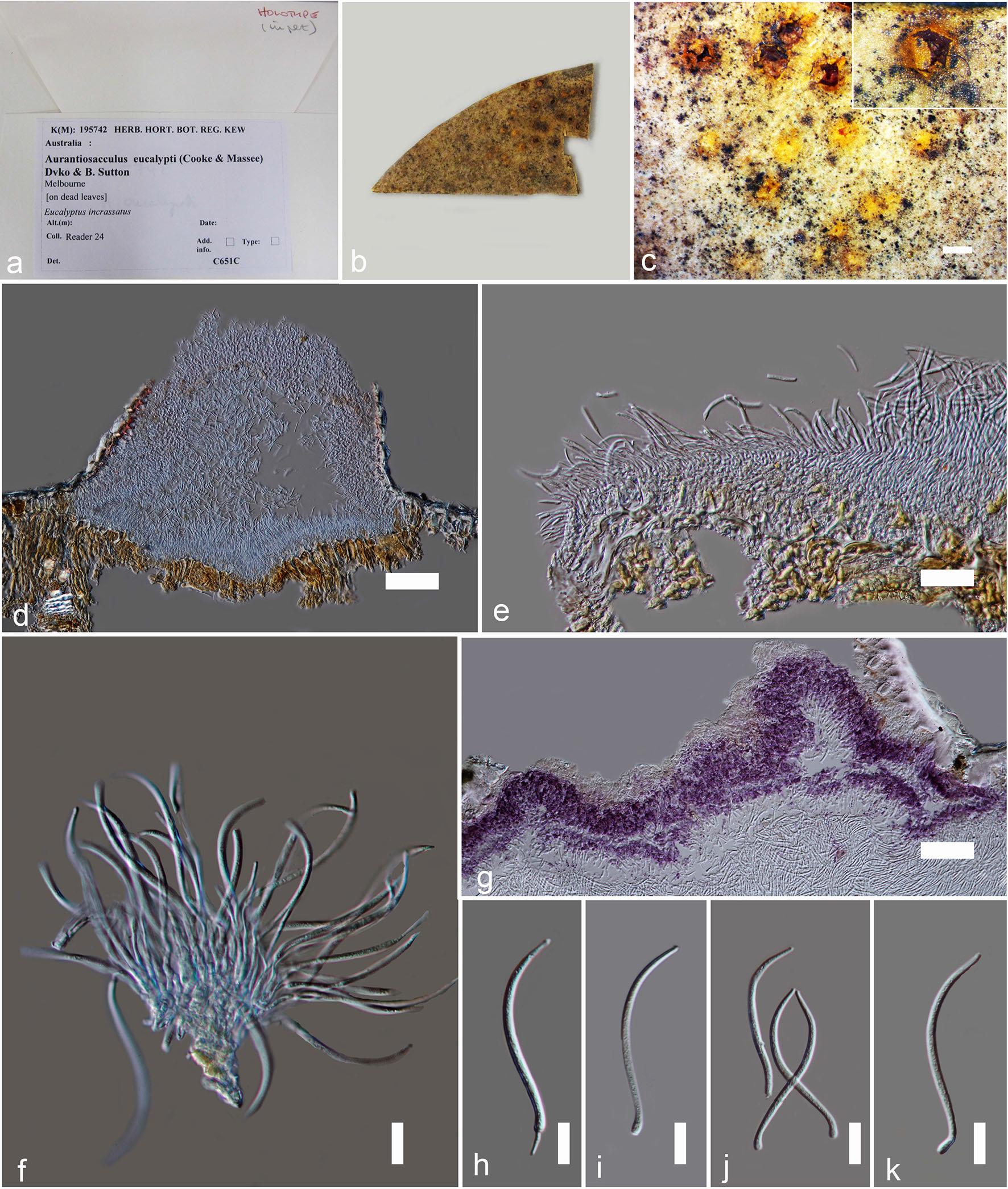
Material examined: AUSTRALIA, Melbourne, on leaves of Eucalyptus incrassatus, Reader 24, holotype K (M) 195742.
Notes: Aurantiosacculus species associated with leaf spots on Eucalyptus baxteri, E. incrassate and E. obliqua (Marshall 1997). Members of this genus only reported from Australia (Crous et al. 2012a). Aurantiosacculus is characterized by having characteristic leaf spots, with bright orange conidiomata with brown furfuraceous tissue and aseptate conidia with swollen bases and thickened scars. Phylogenetic analysis of Crous et al. (2012a) showed the taxonomic placement of Aurantiosacculus within Cryphonectriaceae.
Fig. Aurantiosacculus eucalypti [K (M) 195742]. a Packet of the herbarium. b Herbarium specimen. c Conidiomata on substrate. d Cross section of conidiomata. e Peridium. f Conidia attached to conidiogenesis cells and conidiophores. g Stromatic tissues became purple at KOH. h–k Conidia. Scale bars: d, g = 100 µm , e–f = 20 µm , h–k = 10 µm.