Aposphaeria corallinolutea Gruyter, Aveskamp & Verkley, in Gruyter et al., Stud. Mycol. 75: 28 (2012) [2013]
Index Fungorum number: IF 564798, MycoBank number: MB 564798, FacesofFungi number: FoF 01647, Fig. 28
Saprobic on dead twigs of Prunus padus (Rosaceae) or wood of Fraxinus excelsior (Oleaceae) or undetermined host. Sexual morph: see Tibpromma et al. (2017). Asexual morph: Conidiomata 200–400 µm diam., 180–340 µm high, black, stromatic, pycnidial, solitary to gregarious or confluent, subepidermal, initially immersed to semi-immersed, ultimately become erumpent, round and depressed in surface view, globose to subglobose in section view, unilocular or multilocular, glabrous, ostiolate. Ostiole circular, short papillate, centrally located. Conidiomatal wall 20–40 µm wide, composed of thick-walled, dark brown to brown cells of textura angularis in the periphery, gradually merging with thick-walled, hyaline cells towards conidial hymenium. Conidiophores formed from the inner cavity of the conidiomata are hyaline, cylindrical, branched at the base, septate, smooth-walled. Conidiogenous cells 6–15×1–2.5 µm, hyaline, enteroblastic, phialidic, cylindrical, integrated or discrete, determinate, smooth-walled. Conidia 3–5×1–2 µm (x̄ =3.7×1.4 µm; n=30), hyaline, cylindrical to ellipsoidal, unicellular, smooth, with guttule at each end.
Material examined – Russia, on the dead stem of undetermined host, 27 May 2016, T.S. Bulgakov, DL 5 (MFLU 16-2412)
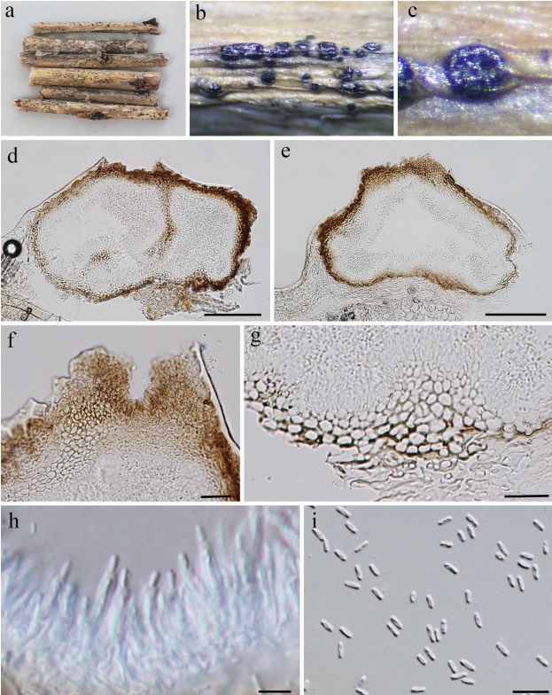
Fig. 1. Aposphaeria corallinolutea (MFLU 16–2412). a Herbarium specimen. b–c Appearance of black conidiomata on the host. d–e Vertical sections of conidiomata. f–g Section of peridium. h Conidiogenous cells and developing conidia. i Conidia. Scale bars d–e=100 µm, f–g=20 µm, h=5 µm, i=10 µm.