Amphisphaeria sorbi Senanayake & K.D. Hyde, in Liu et al., Fungal Diversity: 10.1007/s13225-015-0324-y, [10] (2015)
Index Fungorum number: IF550904, Facesoffungi number: FoF00414
Etymology – Named after the host genus on which the fungus occurs.
Holotypus – MFLU 14–0797
Saprobic on the branch of Sorbus aucuparia L. Sexual morph Ascomata 350–380 μm high × 450–505 μm diam. (x̄ = 370 × 482 μm, n = 10), immersed to erumpent, visible as black spots opening through the cracks of the host surface, solitary, scattered, globose to subglobose, short papillate, ostiole periphysate, dark brown. Peridium 30–35 μm (x̄ = 31 μm, n = 15) at the base, 65–70 μm (x̄ = 68 μm, n = 15) at the neck, comprising 8–10 layers, the inner layer of hyaline cells of textura angularis, the outer layer of brown cells of textura angularis. Paraphyses 2–5 μm wide (x̄ = 3 μm, n = 10), longer than asci, filamentous, septate, embedded in a gelatinous matrix. Asci 125–170 × 9–13 μm (x̄ = 145 × 11 μm, n = 20), 8-spored, unitunicate, cylindrical, short pedicellate, apically rounded, with a J-, apical apparatus. Ascospores 16–24 × 6–8 μm (x̄ = 19 × 6.5 μm, n = 20), uniseriate, rarely overlapping uniseriate, ellipsoidal, light brown, one median septate, slightly constricted at the septum, smooth-walled, surrounded by a thick mucilaginous sheath. Asexual morph Coelomycete. Conidiomata 500–900 μm diam. (x̄ = 800 μm, n = 10), superficial on MEA, solitary or aggregated, globose, dark brown. Peridium consists of thick-walled, septate, brown mycelium. Conidiophores 17–20 μm long, 1.5–2.5 μm (x̄ = 18 × 2 μm, n = 20), arising from peridium, septate, branched, thick-walled, hyaline. Conidiogeneous cell elongated conical, 0.7–1 μm wide at the apex, 2–2.5 μm wide at the base (x̄ = 1 × 2 μm, n = 20), thin-walled, septate, hyaline, annelidic. Conidia 10–12 × 1–1.5 μm (x̄ = 10 × 1 μm, n = 20), elongate-fusiform, hyaline, smooth-walled.
Culture characters – Colonies on MEA reaching 4 cm diam. after 14 days at 18 °C, white, cottony, flat, low, dense, with slightly wavy margin and few aerial mycelia.
Material examined – Italy, Trento [TN], Dimaro, Folgarida, on a branch of Sorbus aucuparia L. (Rosaceae), 2 August 2013, E. Camporesi IT 1400 (MFLU 14–0797, holotype); ex-type living cultures, MFLUCC 13–0721.
GenBank Accession Numbers – LSU: KP744475.
Notes – Amphisphaeria was introduced by Cesati and De Notaris (1863) without designating a generic type (Wang et al 2004). Petrak (1923) proposed A. umbrina as the lectotype of the genus. Different studies have listed more than 250 species in Amphisphaeria, and Wang et al. (2004) accepted 12 species in the genus after examining more than 170 type specimens. Amphisphaeria sorbi shows more similarities to A. vibratilis. Amphisphaeria sorbi, however, differs from A. vibratilis in having small perithecia, a peridium with a cell arrangement of textura angularis, and wide, non-flexuose paraphyses. The ascus apical apparatus is discoid in Amphisphaeria sorbi and has shorter, smooth-walled ascospores without deeply pigmented septa. Molecular analysis of the LSU gene region (Fig. 1) confirms that Amphisphaeria sorbi clusters with A. umbrina in Amphisphaeriaceae with 52 % bootstrap support.
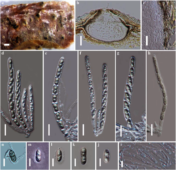
Fig. 1 Amphisphaeria sorbi (holotype). a Ascomata on substrate b Cross section of ascoma c Peridium d–g Asci in water h. Asci inMelzer’s reagent i Paraphyses j–l Ascospores m Sheath around spore n Germinating ascospore. Scale bars: a = 1000 μm, b = 100 μm, c = 50 μm, d–h = 20 μm, i–n = 10 μm.
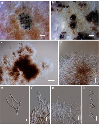
Fig. 2 Amphisphaeria sorbi (ex-type culture) Asexual morph in culture. a, b Conidiomata on MEA. c Peridium. d–g Conidiophore and conidiogenous cells with attached conidia. h Conidia. Scale bars: a =500 μm, b = 1000 μm, c = 100 μm, d–h = 10 μm.