Allophaeosphaeria subcylindrospora W.J. Li, Camporesi & K.D. Hyde.
Index Fungorum number: IF551396; Facesoffungi number: FoF00948; Fig. 2
Etymology – In reference to the subcylindrical conidia.
Holotype – MFLU 15 – 0697
Saprobic on dead stem of Dactylis glomerata L. (Poaceae), forming conspicuous, small, rounded, black fruiting bodies. Sexual morph: Undetermined. Asexual morph: Coelomycetous. Conidiomata 65 – 70 μm high, 83 – 93 μm diam., pycnidial, solitary, gregarious or confluent, subepidermal, immersed to semi – immersed, globose, pale brown to brown, unilocular, ostiolate. Wall of conidiomata 6–11 μm wide, composed of 4–5-layers,thin-walled cells of textura angularis, with cells of outer 1 – 2 layers pale brown, gradually merging with inner 1 – 2 hyaline layers. Ostiole 5 – 7 μm wide, rounded, central , comprising dark brown to black cells . Conidiophores reduced to conidiogenous cells. Conidiogenous cells phialidic, subcylindrical, hyaline, arising from inner region of conidiomata. Conidia 14 – 22 × 1.6 – 3.7 μm (x̄ = 19 × 2.6 μm; n = 30), fusiform to subcylindrical, with an obtuse and narrow apex, and a truncate base, pale brown, mostly 3 – septate, occasionally 1-septate, not constricted at septa, thick -walled, smoothwalled.
Culture characteristics – Colonies on PDA flat, circular, with dull green margin, mouse-grey in the middle and pale mouse-grey at the center; reverse dull green.
Material examined – ITALY, Province of Trento [TN], Val di Sole, Cogolo, on dead stem of Dactylis glomerata (Poaceae), 30 June 2012, Erio Camporesi, IT-520 (MFLU15–0697, holotype); ex-type living culture, MFLUCC 13- 0380, KUMCC 15–0090; ibid. (KUN! HKAS 89500, isotype).
Notes – In the present paper, we introduce an asexual morph taxon, A. subcylindrospora, which is phylogenetically close to, but distinct from A. muriformia (Fig.1). Allophaeosphaeria lacks any known asexual morph (Liu et al. 2015) . Allophaeosphaeria subcylindrospora resembles P. papayae (Speg.) Quaedvl. et al. in having pycnidial, brown, globose conidiomata and subcylindrical, pale brown conidia with an obtuse apex, and truncate base. However, A. subcylindrospora is further characterized by cylindrical conidiogenous cells, and mostly 3 – septate, occasionally 1 – septate conidia, which are shorter than P. papaya (15–) 26 – 32 (−35) × (2.5–) 3 μm (Quaedvlieg et al. 2013).
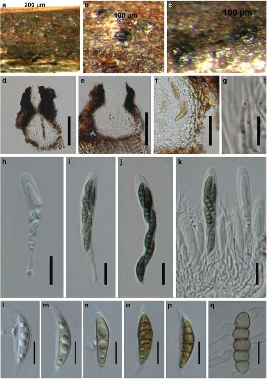
Fig.1 Lophiostoma caulium (reference specimen) a–c Appearance of immersed ascomata on host surface d, e Hand sections of ascomata. Note the ostiole, with a small to large, flat, crest-like apex f Peridium g. Hamathecium h – k Asci with ascospores l – p Ascospores o. Germinating ascospore. Scale bars: d, e = 100 μm, f, h – k = 20 μm, g = 10 μm, l – q = 10 μm.
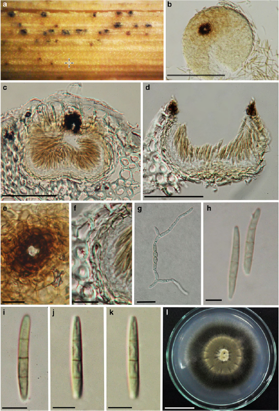
Fig. 2 Allophaeosphaeria subcylindrospora (holotype) a Specimen showing appearance of taxon on host b Surface view of conidioma c, d Vertical section of conidioma with conidiogenous cells and developing conidia e Surface view of ostiole f Section of peridium g Germinating conidia h – k Conidia l Culture on PDA. Scale bars: b = 100 μm, c – d = 50 μm, e – f = 10 μm, g = 20 μm, h, i, k = 5 μm, l = 25 μm.