Allophaeosphaeria muriformia Ariyawansa, Camporesi & K.D. Hyde.
Index Fungorum number: IF550998, Facesoffunginumber: FoF00410; Fig. 1
Etymology – The specific epithet muriformia is based on the ascospore septation.
Holotype – MFLU 15-0067
Saprobic on dead wood. Sexual morph Ascomata 275 – 340 μm high × 300 – 380 μm diam. (x̄ = 290 × 340 μm), solitary, scattered, superficial, globose to subglobose, ostiolate. Ostiole papillate, black, smooth, with neck and without periphyses Peridium 45 – 70 μm wide, comprising 2 – layers, outer layer composed of heavily pigmented thick-walled cells, innermost layer of broad, hyaline compressed rows of cells of textura angularis. Hamathecium lacking pseudoparaphyses. Asci 210 – 237 × 50 – 70 μm (x̄ = 235 × 62 μm, n = 20), 8 – spored, bitunicate, fissitunicate, elongate cylindrical to slightly clavate, with a minute pedicel, thick-walled and rounded at apex, with an ocular chamber. Ascospores 40 – 60 × 20 – 30 μm (x̄ = 56 × 26 μm, n = 40), overlapping 2 – 3 – seriate, oblong to narrowly oblong, straight to slightly curved, muriform, multi-septate, constricted at each septa, hyaline, pale brown when mature, smooth – walled. Asexual morph Undetermined.
Material examined – ITALY, Province of Forlì-Cesena, Montevescovo, on dead stem, 4 February 2013, E.Camporesi (MFLU 14–1122, holotype), ex-type living culture, MFLUCC 13–0349. GenBank ITS: KP765680; LSU: KP765681; SSU: KP765682.
Notes – Allophaeosphaeria resembles many species of Phaeosphaeria in having a peridium comprising 2 – 3 layers of brown to dark brown cells of textura angularis and multiseptate ascospores with a gelatinous sheath, but differs in having elongate-cylindrical to slightly clavate asci with a clear ocular chamber and lacks pseudoparaphyses. The phylogenetic analysis of combined ITS, LSU and SSU sequences provided strong evidence that the type species of Allophaeosphaeria, A. muriformia belongs in Phaeosphaeriaceae and clustered together with putatively named strains of Phaeosphaeria phragmiticola (CBS 459.84) and Phaeosphaeria vagans (CBS 604.86).
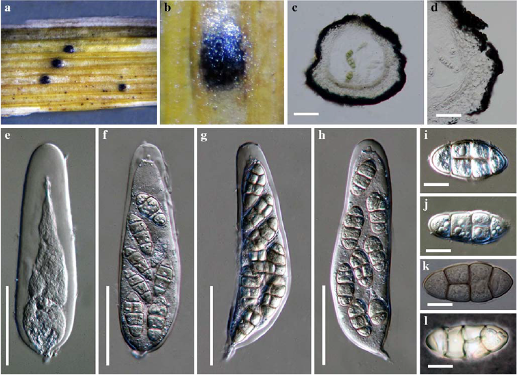
Fig. 1 Allophaeosphaeria muriformia (holotype) a, b Ascomata on the host surface c Section of an ascoma d Close up of the peridium e–h Cylindrical asci with a minute pedicel i–k Hyaline to pale brown ascospores k Ascospores mounted in Indian ink. Scale bars: c = 50 μm, d = 5 μm, e – g = 30 μm, h – j = 5 μm, k = 10 μm.