Allantophomopsis fusiformis (Nag Raj) Nag Raj, Coelomycetous Anamorphs with Appendage-bearing Conidia: 114 (1993)
≡ Apostrasseria fusiformis Nag Raj, Can. J. Bot. 61: 17 (1983)
= Phacidium taxicola Dearn. & House, Mycologia 18(5): 239 (1926)
Index Fungorum Number: IF 359573; Facesoffungi number: FoF 07106
Saprobic on leaves and stems of Taxus canadensis. Sexual morph: Apothecia 290–380 μm diam., 130–300 μm high, black at the base, brown at the upper part in outline, solitary to gregarious, or confluent, subepidermal, initially immersed to semi-immersed, ultimately becoming erumpent, ellipsoid to conical, unilocular, glabrous, ostiole absent, dehiscing by an irregular split in the middle part of overlapping host tissue. Peridium 10–45 μm wide, composed of thick-walled, brown to dark brown cells of textura angularis. Hamathecium comprising paraphyses and asci. Paraphyses 30–55 × 1–3 μm, hyaline, numerous, slender, with an obtuse apex, branched and slightly swollen at the base, anastomosing, septate, often constrict at septa. Asci 34–49 × 4–7 μm ( ̄x = 41 × 5.6 μm; n = 30), unitunicate, 8-spored, clavate, bluntly rounded at apex, with a distinct apical ring, short-pedicellate, with an amyloid (J+) apical apparatus. Ascospores 7–10 × 2–3.5 μm ( ̄x = 8.5 × 3 μm; n = 30), hyaline uniseriate or biseriate, or irregularly arranged, ellipsoid to fusiform, acute at both ends, unicellular, smooth-walled, guttulate. Asexual morph: Conidiomata 170–250 μm diam., 140–200 μm high, brown to dark brown, stromatic, pycnidial, solitary to gregarious, developing in linear series on both sides of the midrib, appearing as slightly elevated, minute, brown to black, rounded specks in surface view, sub-peridermal, deeply immersed, globose to subglobose, unilocular or irregularly plurilocular, glabrous, papillate, ostiolate. Ostiole 50–70 × 40–55 μm, single. cylindrical to circular, centrally located. Conidiomatal wall 10–30 μm wide, composed of thick-walled, pale brown to hyaline cells of textura angularis to textura prismatica in the basal and lateral part, passing into thick-walled, hyaline cells of textura prismatica in the locular wall, becoming olivaceous-brown cells of textura angularis at ostiolar region. Conidiophores formed from the innermost layers of conidiomata, hyaline, subcylindrical, branched at base, septate, often constricted at septa, smooth-walled. Conidiogenous cells 4–10 × 2–4 μm, hyaline, enteroblastic, annellidic, with 1–3 annellations, cylindrical to ampulliform, discrete or integrated, smooth-walled. Conidia 6–9.5 × 2–3.5 μm ( ̄x = 8 × 3 μm, n = 30), hyaline, fusiform to occasionally naviculate, with an acute apex and a
narrow truncate base, unicellular, smooth-walled, guttulate, with a funnel-shaped or irregular, mucoid, apical appendage, sometimes with a short, less conspicuous, mucoid, basal appendage.
Material examined – USA, New York, Hamilton Co., Blue Mountain Lake, on leaves and stems of Taxus canadensis (Taxaceae), 7 September 1920, H.D. House (JD 4882, type).
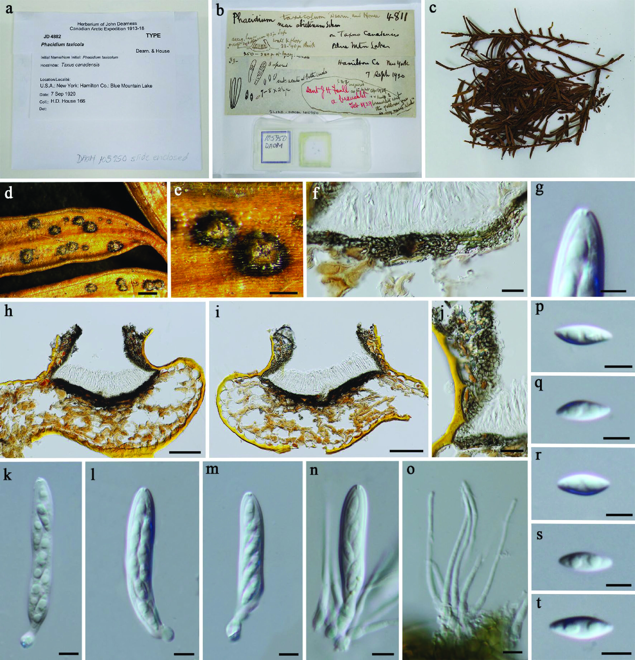
Fig. 1. Allantophomopsis fusiformis (sexual morph, JD 4882, type). a–c Herbarium package and specimen. d–e Appearance of black apothecia on the host. f, j Section of peridium. g Enlarged view of apical ascus. h–i Vertical sections of apothecia. k–n Asci. o Paraphyses. p–t Ascospores. Scale bars d = 500 μm, e = 200 μm, f, j = 20 μm, g, k–t = 5 μm, h–i = 100 μm.
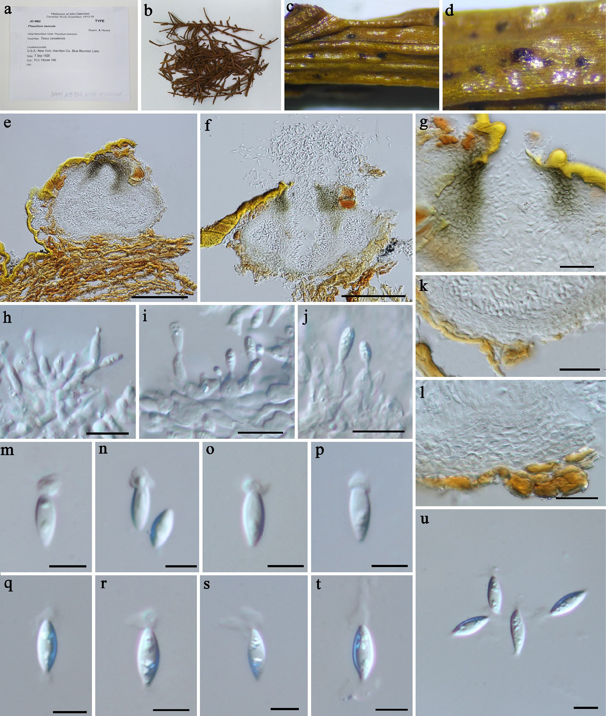
Fig. 2. Allantophomopsiella pseudotsugae (DAOM 129883). a, b Herbarium package and specimen. c–e Appearance of dark brown to black conidiomata on the host. f Ostiole. g Vertical section of conidiomatal wall in ostiolar region. h–j Vertical sections of conidiomata. k–n Conidiophores, conidiogenous cells and developing conidia. o–t Conidia. Scale bars: c–d = 500 μm, e = 200 μm, f–g = 50 μm, h–j = 200 μm, k–m = 10 μm, n–t = 5 μm.