Allantophomopsiella pseudotsugae (M. Wilson) Crous, in Crous, Quaedvlieg, Hansen, Hawksworth & Groenewald, IMA Fungus 5(1): 180 (2014)
≡ Phomopsis pseudotsugae M. Wilson, Trans. R. Scottish Arboricult. Soc. 34(2): 147 (1920)
= Allantophomopsis pseudotsugae (M. Wilson) Nag Raj, Coelomycetous Anamorphs with Appendage-bearing Conidia: 116 (1993)
= Potebniamyces coniferarum (G.G. Hahn) Smerlis, Can. J. Bot. 40: 352 (1962)
Index Fungorum Number: IF 809674;Facesoffungi number: FoF 07104
Saprobic on branches of Pinus sylvestris. Sexual morph: see DiCosmo et al. (1983). Asexual morph: Mycelium immersed, composed of septate, branched, hyphae. Conidiomata 260–570 μm diam., 300–500 μm high, dark brown to black, stromatic, pycnidial, solitary to gregarious, or confluent, initially immersed, ultimately becoming erumpent, pulvinate to subconical, irregularly plurilocular, thick-walled, glabrous, papillate, ostiolate. Ostiole 60–90 × 60–70 μm, single. short-cylindrical, centrally located. Conidiomatal wall 20–40 μm wide, composed of thick-walled, dark brown to brown cells of textura epidermoidea to textura intricata in the basal and lateral part, passing into thick-walled, pale brown cells of textura epidermoidea in locular wall, with dark brown cells of texture intricata at ostiolar region. Conidiophores formed from the innermost layers of conidiomata, hyaline, subcylindrical, branched or unbranched, septate, often constricted at septa, smooth-walled, invested in mucus. Conidiogenous cells 5–9 × 2–4 μm, hyaline, enteroblastic, annellidic, lageniform to subcylindrical, discrete or most often integrated, determined, smooth-walled, with new subtending cells becoming conidiogenous cells. Conidia 5–8 × 2–3 μm ( ̄x = 6 × 2.5, n = 30) μm, hyaline, ellipsoid, obovoid or occasionally fusiform, obtuse or acute at apex, narrow and truncate at base, unicellular, thick and smooth-walled, guttulate, bearing a flame-like or irregular, mucoid apical appendage and sometimes, a shorter and less conspicuous, mucoid, basal appendage.
Material examined – Canada, Quebec, Lac-à-la-Tortue, on branches of Pinus sylvestris (Pinaceae), 10 November 1967, E. Smerlis (DAOM 129883).
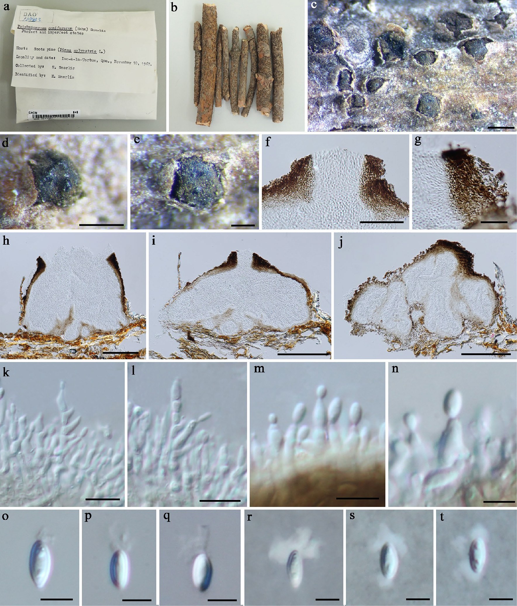
Figure 1. Allantophomopsiella pseudotsugae (DAOM 129883) a, b Herbarium package and specimen. c–e Appearance of dark brown to black conidiomata on the host. f Ostiole. g Vertical section of conidiomatal wall in ostiolar region. h–j Vertical sections of conidiomata. k–n Conidiophores, conidiogenous cells and developing conidia. o–t Conidia. Scale bars: c–d = 500 μm, e = 200 μm, f–g = 50 μm, h–j = 200 μm, k–m = 10 μm, n–t = 5 μm.