Acrospermum urticae D. Pem, Camporesi & K.D. Hyde, sp. nov.
MycoBank number: MB 556687; Index Fungorum number: IF 556687; Facesoffungi number: FoF 06382; Fig. 4
Etymology: Name reflects the host from which the fungus is isolated.
Holotype: MFLU 18-1666.
Saprobic on dead stem of Urtica dioica. Sexual morph: Ascomata 940–1057 high × 301–345 µm diam. ( x̄ = 1021 × 318 µm), solitary or in groups, superficial, club-shaped to conoid, erect, unilocular, brown to blackish when dry, with a short stipe or sessile, flattened when dry, swelling when moist, ostiole large, apex rounded. Peridium 11–12 µm in vertical section comprising three layers, an outer layer comprising dark brown cells of textura angularis, a central thick layer, comprising pale brown to hyaline tissue of gelatinized hyphae with elongated cells, and an inner layer comprising dense tissue of small, hyaline cells. Hamathecium comprising narrow, long, pseudoparaphyses. Asci 195–319 × 6.2–6.7 μm ( x̄ = 252.5 × 6.4 µm), 8-spored, bitunicate, narrowly cylindrical, pedicellate, with an ocular chamber. Ascospores 122–170 × 1.1–1.2 μm ( x̄ = 146.2 × 1.2 µm), fasciculate, filiform, hyaline, multi-septate, nearly as long as the asci, smooth-walled. Asexual morph: undetermined
Material examined: Italy, Ravenna [RA], San Cassiano di Brisighella, on dead aerial stem of Urtica dioica (Urticaceae), 13 August 2018, Erio Camporesi (IT 3999, holotype; MFLU 18-1666, isotype).
GenBank numbers: LSU: MN597994, SSU: MN597996.
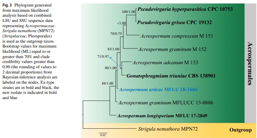
Notes: Acrospermum urticae differs from Acrospermum longisporium by its smaller ascomata (940–1057 high × 301–345 µm diam. v.s. 1500–2000 high × 400–500 µm diam.) and wider ascospores (122.1–175.3 × 1.1–1.2 μm v.s. 150–170 × 0.5–1 μm). Phylogenetic analyses of a combined LSU, SSU sequence dataset show that A. urticae forms a distinct lineage in Acrospermaceae with strong ML and BYPP support (80% ML, 1.0 BYPP; Fig. 3). Therefore, we introduce Acrospermum urticae as a new species.


Fig. 4 Acrospermum urticae (IT 3999, holotype). a–d Ascomata on host surface. e Ascoma in vertical section f Peridium. g–i Narrowly cylindrical asci. j–k Filiform ascospores Scale bars: a = 2000 µm, b, e = 500 µm, c, d = 300 µm, f, g = 100 µm, h = 25 µm, i = 30 µm, j = 50 µm, k = 40 µm
Species
