Ionopezia gerardii (Cooke) Matočec, I. Kušan & Jadan, comb. nov.
Index Fungorum number: IF 556252; MycoBank number: MB 556252; Facesoffungi number: FoF 07862; Figs. 42, 43
Basionym: Peziza gerardii Cooke, Hedwigia 14(6): 81 (1875)
= Peziza ionella Quél., Bull. Soc. Bot. Fr. 24: 328 (1878) [1877]
= Plicaria pedicellata Velen., Novitates Mycol.: 198 (1940)
For complete synonymy see Pfister (1978) and Van Vooren (2009).
Ascomata apothecial, individual, circular from the top view, larger fruitbodies can be slightly irregular from the top view, cupulate when young, then shallowly saucer-shaped to plate-shaped, short stipitate, 3.8–6.8 mm diam, hymenial surface greyish violet to vividly violet, matte, not wrinkled, margin upright at first, then flattened, beset with whitish floccose granules on pale greyish violet ground, granules up to 0.5 mm tall; excipulum surface concolourous with the margin, beset with less floccose granules; on section hymenium creamy grey, subhymenium differentiated due to dark violet pigmentation, flesh creamy grey, not exuding latex, marginal and excipular flank granules whitish and conical, stipe-like base 1.9–2.1 mm wide, 0.5–0.7 mm high, without pronounced subiculum. Hymenium 330–380 µm thick, arranged as a regular palisade. Subhymenium 70–90 µm thick, composed of textura angularis-epidermoidea, cells 5.1–15.7 µm wide, thin-walled and hyaline, but richly beset by dark violet–purple pigment, cells often contain one to few strongly refractive globules, due to the high concentration of pigment well discerned from the medulla. Marginal texture differentiated into perihymenial area, extreme margin/proximal margin as a whole, distal margin and marginal medulla: (1) perihymenial area wholly compact, composed of narrowly prismatic cells arranged as textura porrecta with thin and hyaline cell walls, encrusted with dark purple to violet plaques and intercellular diffuse purplish matrix, especially on a surface, cells 3.5–7.8 µm wide, this area is rather unclearly distinguished from; (2) proximal margin, composed of wider prismatic cells forming textura prismatica oriented perpendicularly to the surface with protruding individual elements, cells are thin-walled and hyaline, with less pigments and more abundant intercellular matrix, cells 5.6–12.2 µm wide, terminal cells often clavate, these cell chains arising from basal; (3) marginal medulla textura angularis with cells 6.5–16.2 µm wide, hyaline and thin-walled but fairly rich in purple-violet plaques and diffuse pale purple pigment in intercellular spaces; (4) extreme margin formed as a single pustule, 75–130 µm wide and 18–74 µm high, composed entirely as proximal margin; (5) distal margin of 2–4 pustules, 75–115 µm wide, 15–70 µm high, composed of hyaline and thin-walled shortly prismatic cells to ellipsoid to clavate ones, rather untidily organized, cells 12.4–29.1 × 7.2–17.4 µm, entirely without any refractive/pigment inclusions, cell chains in distal marginal pustules originate from basal marginal medullar textura angularis with cells 7–11.2 µm wide in deeper areas and 8–18 µm wide in the area immediately below the pustules, this area is very rich in both plaque-form darker consolidated pigments as well in diffuse ones. Medullary excipulum unclearly differentiated into two sublayers, altogether 212–260 µm thick, upper part 160–185 µm thick, composed of textura ellipsoidea-epidermoidea, cells 5–22.3 µm, mostly thin-walled and hyaline, some individual cells thick-walled, without intracellular refractive/pigment inclusions but rich in diffuse pale violet–purple intercellular matrix, lower part 38–75 µm thick, composed of textura intricata-porrecta to intricata-prismatica with wavy cell chains running ± parallel with the surface, hyphae 4–12.5 µm wide, cells thin-walled, hyaline, principally without any pigment/refractive intra- or intercellular content. Ectal excipulum composed of weakly developed discontinuous cortex and thick continuous layer; discontinuous part composed of cell aggregates of textura globulosa-subangularis, 30–95 µm high, 47–105 µm wide, individual cells globose, subangular to shortly clavate, 8.8–22.5 µm wide, walls hyaline, somewhat thickened, 0.7–2 µm thick, without any internal refractive/pigment inclusions but with apparent hyaline intercellular matrix; continuous part 130–173 µm thick, composed of textura angularis-prismatica, cell chains directed ± perpendicularly to the surface, cells ellipsoid, clavate to prismatic, 16–86.5 × 6.4–37.8 µm, hyaline, thin-walled, intercellular spaces with some hyaline matrix only in outermost area, refractive/pigment inclusions absent. In Cotton Blue overall texture not cyanophilous; in Lugol's solution terminal cells of ectal excipulum may contain Lugol-induced bodies; in KOH pigmented granules and ectochroic plaques soluble; in HCl inert. Paraphyses cylindrical, apically obtuse, ± straight, not branching in the upper part, apical cell 28.4–110 × 4.5–7.5 µm, containing few weakly refractive globules, embedded in subhyaline to faintly violet, sparse and faintly developed exudate, containing also strongly refractive granules/plaques, not forming continuous layer; Lugol's solution causes formation of refractive sienna purple to brownish purple granules in paraphysis cytoplasm. Asci 265–345 × 16.4–18 µm, cylindrical to slightly ventricose, broadest point is in a lowest zone of pars sporifera, apex subtruncate-rounded, protruding above paraphysis tips up to 22.5 µm at full maturity, pars sporifera 97–108 µm, 8-spored, base tapered, pleurorhynchous, arising from croziers, operculum only faintly predelimited, 4.3–4.6 µm diam, 0.7–0.8 µm thick, strictly apical, flat-lenticular, encircled by rather broad but very shallow indentation ring, 1–1.1 µm wide, periascal mucus thin, film-like, evenly thick throughout, 1–2 perimarginal asci contain at least lower 4 globose to subglobose spores; in Lugol's solution periascal mucus amyloid, reaction of medium strength in upper ascus area, intensity gradually decreasing towards the base. Ascospores (20–)21.1–26–27.9(–28.5) × 7.5–8.9–11(–11.2) µm, Q = 1.98–3.02–3.45(–3.60) (n = 200, in H2O), hyaline, fusiform to naviform, with subacute poles, even slightly papillate, slightly asymmetrical, 1-celled, smooth under immersion lens when in water mount, in Cotton Blue elongated striae visible but not cyanophilic, multiguttulate, with 2–3 large lipid bodies, 3.2–6.2 µm diam, and many smaller ones, 0.7–2.8 µm diam, uninucleate, nucleus 2.1–2.3 µm diam, equatorially-peritunically positioned, thin-walled, wall 0.5 µm thick, freshly ejected without sheath; in Cotton Blue without de Bary bubbles, perispore not cyanophilic or not present.
Habitat and phenology: Terricolous, directly on forest soil, sometimes with mosses, on river banks and pathway edges, inhabiting biomes from colline to arcto-alpine zone of Northern Hemisphere, fruitbodies appear from spring to autumn.
Known distribution: The species is known from North America, Europe, and India.
Material examined: CROATIA, Zagreb City, Park forest Maksimir, 45°49′12″ N, 16°00′59″ E, 120 m a.s.l., Pinus sylvestris plantation, directly on forest soil, 25 May 1989, N. Matočec (CNF 2/947); Park forest Jelenovac, 45°49′32″ N, 15°57′03″ E, 170 m a.s.l., forest of Quercus petraea and Carpinus betulus, directly on moist soil near the creek, 14 July 1990, N. Matočec, (CNF 2/1301); Park forest Maksimir, 170 m NE from Vidikovac, 45°49′33″ N, 16°01′23″ E, 135 m a.s.l., forest of Quercus petraea, Carpinus betulus, on forest soil with litter, 12 October 2005, N. Matočec and I. Kušan (CNF 2/7450); Medvednica mountain, vicinity of Kraljičin zdenac hut, 600 m SE from Medvedgrad castle, 45°51′59″ N, 15°56′49″ E, 370 m a.s.l., forest of Fagus sylvatica, Quercus petraea, Carpinus betulus, Castanea sativa, on forest soil at the edge of a path, 14 July 2013, D. Mrvoš and N. Matočec (CNF 2/9431); Virovitica-Podravina County, Dumače area in Bilogora range, 9.1 km SW – S-SW from Virovitica, 45°45′55″ N, 17°19′19″ E, 180 m a.s.l., forest of Fagus sylvatica, Tilia sp. and Acer sp., forest soil near the creek, 15 August 1997, S. Gottstein (CNF 2/3493); BOSNIA AND HERZEGOVINA, Sarajevo County, Igman mountain plateau, Javornik area, 43°44′54″ N, 18°14′04″ E, 1480 m a.s.l., forest of Picea abies, Abies alba, Fagus sylvatica, Acer pseudoplatanus, on forest soil at path edge near creek, 10 August 2018, N. Jukić, I. Kušan and N. Matočec (CNF 2/10798, duplex FAMU N.J. 100818-Y9).
GenBank numbers: ITS: MK514543; LSU: MK514537; EF1-α: MK705937; RPB2: MK673768 (CNF 2/10798).
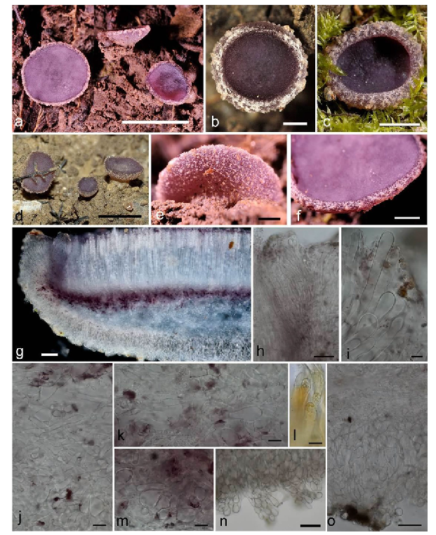
Fig. 42 Ionopezia gerardii. a-f Ascomata. g Vertical median section through apothecium. h Perihymenium and proximal margin. i Terminal cells on extreme margin. j Medullary excipulum. k Contact zone between subhymenium and medullary excipulum. l Terminal cells of ectal excipulum with Lugol induced bodies. m Subhymenium. n Cell aggregates on ectal excipulum. o Ectal excipulum. g–k, m–o In water mount (*), l In Lugol's solution (*). g In dark field, a –f, h–o In bright field. a, e, f–o From CNF 2/10798, b, d From CNF 2/7450, c From CNF 2/9431. Photo: N. Matočec and I. Kušan. Scale bars: a, d = 5 mm, c = 2 mm, b, e, f = 1 mm, g = 100 µm, h, n, o = 50 µm, i–m = 10 µm. Asterisk (*) denotes living material
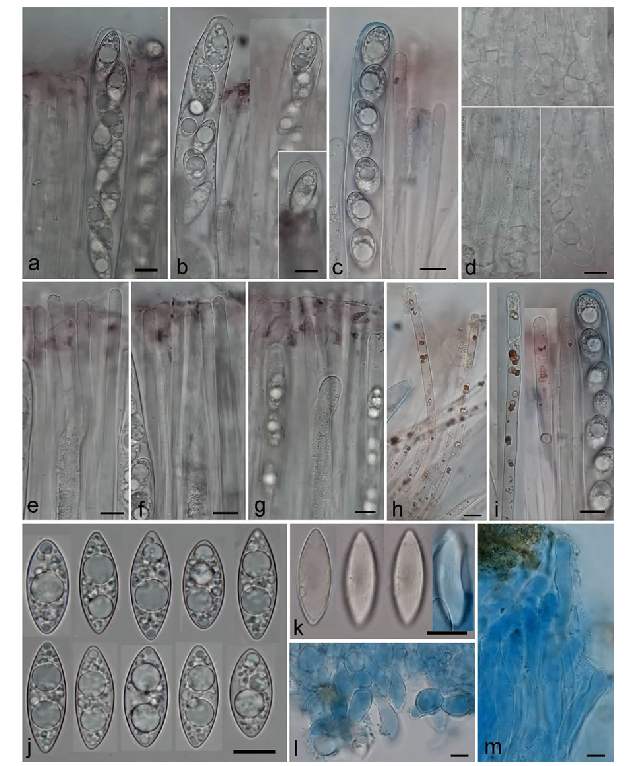
Fig. 43 Ionopezia gerardii. a–c Living mature asci with paraphyses. d Ascus bases with ascogenous cells. e–g Paraphyses with epithecial gel. h, i Paraphyses with Lugol induced granules. j, k Ascospores. l Terminal cells on ectal excipulum. m Margin. a, b, d–g, j In water mount (*), c, h, i In Lugol's solution (*), k-m In Cotton Blue (†). All from CNF 2/10798. Photo: N. Matočec and I. Kušan. Scale bars: a–m = 10 µm. Asterisk (*) denotes living material. Cross (†) denotes dead material