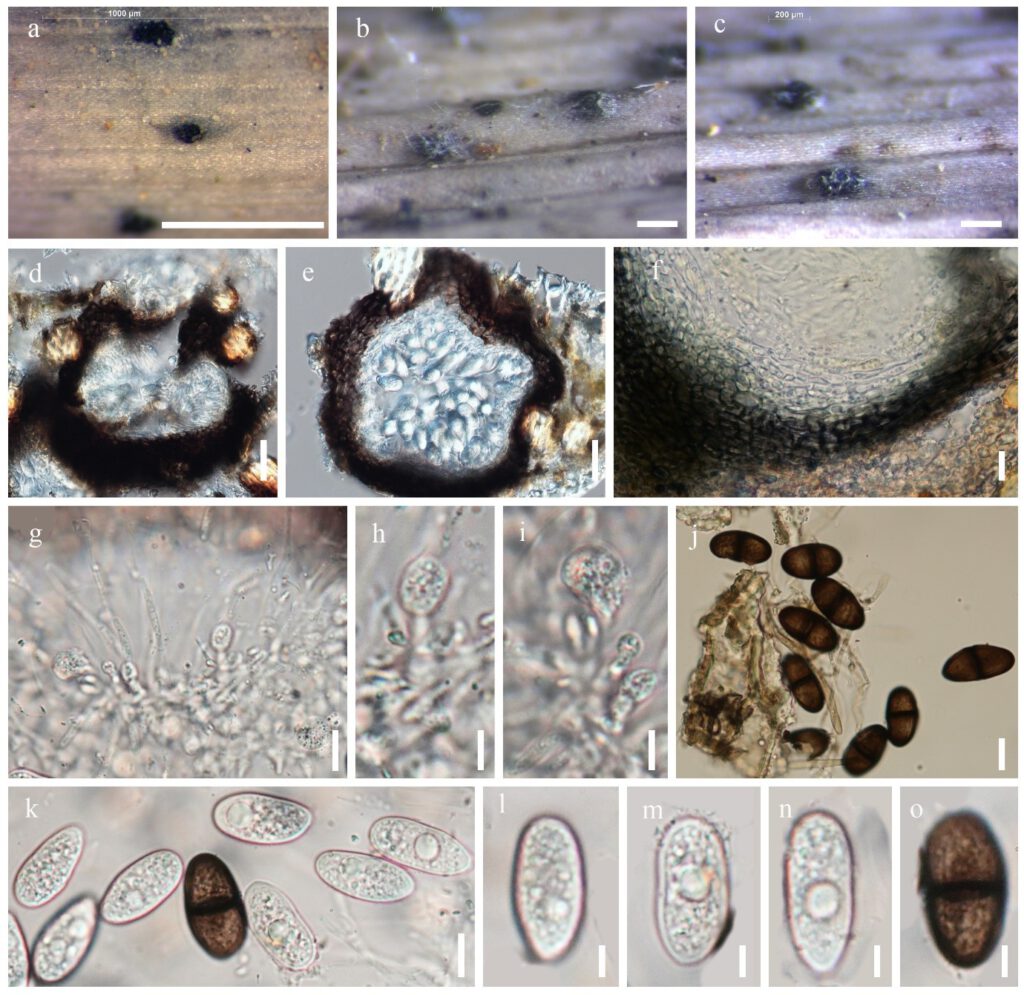
Lasiodiplodia pseudotheobromae A.J.L. Phillips et al., Fungal Diversity 28: 8 (2008) Fig. **
MycoBank number: MB 510941; Index Fungorum number: IF 510941; Facesoffungi number: FoF 00166;
Saprobic on dead leaves of Dracaena fragrans. Sexual morph: Undetermined. Asexual morph: Conidiomata 200–300 μm high × 150–250 μm diam. (x̅= 250×200 μm n = 10), pycnidial, semi-immersed, unilocular, solitary, scattered, globose or subglobose, dark brown. Conidiomata wall 50–65 μm wide, outer layers dark brown to black, thick walled, inner layers thin-walled, pale brown to hyaline, comprising 2–3 layers of dark brown cells of textura angularis. Paraphyses up to 40 μm long, 1.5–2.5 μm wide, hyaline, septate, cylindrical, occasionally branched, ends rounded. Conidiogenous cells holoblastic, hyaline, thin-walled, smooth, cylindrical. Conidiogenous cells 19–33× 5–10 μm (x̅ = 26 ×7.5 μm n= 10), holoblastic, hyaline, cylindrical. Conidia 30–50 μm×10–15 μm (x̅ = 40×12.5 μm n=30), initially hyaline and aseptate when immature, becoming medianly one septate, dark brown, thick-walled, ellipsoid to obovoid, base truncate or rounded, with longitudinal striations from apex to base.
Material examined – THAILAND, Kanchanaburi Province, Sangkhla Buri District, on dead leaves of Dracaena fragrans, 24 October 2018, Napalai Chaiwan, KAN3, MFLU 1***–***, KLV2, MFLU 1***–***, living culture MFLUCC 12–****.
Host – Known to inhabit fruits, leaves and stems of multiple plant genera in multiple families (Doilom et al. 2015, 2016, Pipattanapuckdee et al. 2019, Li et al. 2020) and dead leaves of Dracaena fragrans (This study).
Distribution – Widely distributed in tropical and subtropical regions (Doilom et al. 2015, 2016, Pipattanapuckdee et al. 2019, Li et al. 2020).
GenBank accession numbers – KAN3; ITS: ***. KLV2; ITS: ***, RPB2: ***, TEF1:***, TUB: ***
Notes – The morphology of this collection is similar to L. pseudotheobromae (Gomdola et al. 2020). It possesses septate paraphyses, the conidia are slightly bigger and larger (22–33 × 13–15 versus 30–50 × 10–15 μm) (Pipattanapuckdee et al. 2019, Gomdola et al. 2020). This may be due to the difference in the substrate. However, in the phylogenetic analyses of L. pseudotheobromae isolates from this study grouped it with L. pseudotheobromae species with 67% ML. Thus we identified the species as L. pseudotheobromae. This is the first report of L. pseudotheobromae from Dracaena fragrans in Thailand.

Figure***– Lasiodiplodia pseudotheobromae (KAN3) a–c. Conidiomata on dead leaves of Dracaena fragrans. d–e. Section through conidioma f. Peridium (Conidioma wall) g–i. Conidiogenous cells j–o. Conidia and mature conidia Scale bars: a=1000 μm. b=200 μm. d–g, j–o =10 μm. h–i=5 μm.
