Zygosporium masonii S. Hughes, Mycol. Pap. 44: 15 (1951)
Index Fungorum number: IF308038, Facesoffungi number: FoF 06496
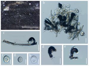
Saprobic on the leaves of Dracaena sp. Asexual morph: Hyphomycetous. Colonies effuse to compact, forming a thin layer, spreading on the substrate surface, numerous, conspicuous, black with flossy mycelium. Mycelium mostly superficial, consisting of cylindrical, light brown to brown, smooth, septate, branched hyphae with slightly thick walls. Conidiophores setiform portion 50–90 × 2–5 μm (x̅ = 66 × 3.5 μm, n = 20), macronematous, mononematous, scattered, up to 7 μm wide near the base, 2–4-septate, erect, straight or slightly flexuous, unbranched, brown at the base with connected conidiophores, subhyaline to light brown and narrowing towards the apex; swollen vesicles on short stalk arising from the side of the first cell of the conidiophores, cylindrical, brown, curved and smooth-walled. Conidiogenous cells 15–30 × 3–7 μm (x̅ = 21 × 5.5μm, n = 10), phialidic, 2–3 per vesicle, ellipsoidal to ampulliform, upwardly curved, smooth, brown to black, apex obtuse, thin-walled, borne in pairs, arising directly from the vesicular cell. Conidia 4–8 × 4–10 μm (x̅ = 6.9 × 6.7 μm, n = 20), solitary, globose to subglobose, aseptate, verruculose, subhyaline to pale brown, granules, thick and rough-walled. Sexual morph: Undetermined.
Material examined – Thailand, Songkhla Province, on dead leaves of Dracaena sp., 5 May 2018, Napalai Chaiwan NSW5 (MFLU 18-0124).
GenBank accession numbers – ITS: MN480499, LSU: MN480500.
Known distribution (based on molecular data) – Australia (Paulus et al. 2007), Cuba (Delgado-Rodriguez et al. 2002), Florida (Delgado 2008), West Indies (Minter 2001), Italy (Lunghini et al. 2013), Japan (Kobayashi 2007), Myanmar (Thaung 2008), Nicaragua (Delgado 2011), Thailand (this study)
Known hosts (based on molecular data) – Artabotrys burmanicus (Thaung 2008), Cecropia peltata (Minter 2001), Daphniphyllum macropodum (Kobayashi 2007), Ficus pleurocarpa (Paulus et al. 2007), Cyathea sp. (Delgado-Rodriguez et al. 2002), Phillyrea angustifolia (Lunghini et al. 2013), Roystonea sp. (Delgado 2011), Tillandsia sp. (Delgado 2008), Dracaena (this study).
Notes – Based on the multigene analyses, our isolate clustered with Zygosporium masonii with high boostrap support (Fig. 82). Zygosporium masonii differs from our isolate in having ovoid, hyaline conidia (Whitton et al. 2012), while our isolate has subglobose, subhyaline to pale brown conidia. The Blastn search results showed that the ITS and LSU sequences of our isolate are 100% identical to those of Z. masonii isolates (CBS 557.73 and CBS 138.71).

Fig. 1 – Zygosporium masonii (MFLU 18–0124, new geographical and host record). a Appearance on host surface. b–e Vesicle, conidiophores and conidiogenous cell. f–h conidia. Scale bars: a = 1000 µm; b, c = 50 μm; e, d, e = 20 μm; f, g, h = 10 μm.
