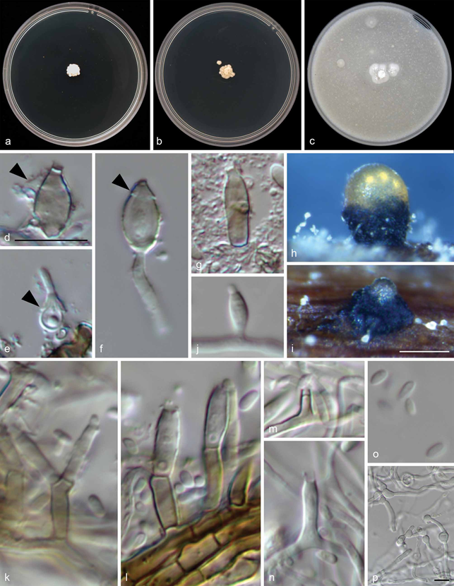
Vredendaliella oleae C.F.J. Spies, Moyo, Halleen & L. Mostert, sp. nov.
MycoBank number: MB 836263; Index Fungorum number: IF 836263; Facesoffungi number: FoF; Fig. 6
Etymology. Referring to the host from which this species was recovered.
Mycelium smooth-walled, forming irregularly swollen hyphal cells on PDA, hyaline, sometimes dark brown, 1– 2.5 (av. 1.5) μm. Conidia forming on hyphal cells and in pycnidia. Pycnidia forming on pine needles on SNA after 4 wk, globose to irregularly globose (50 –)60.5 –160.5(–170.5) (av. 106.5) μm. Seemingly opening by irregular rupture to exude clear conidial suspension. Conidiogenous cells in pycnidia usually dark brown, ellipsoidal to broadly ellipsoidal, oval or lens-shaped, sometimes ampulliform, fusiform or cylindrical, often with bib- like collar, (4.5 –)5 –11(–12.5) × 2 – 5 (av. 7.5 × 4) μm; collarettes inconspicuous, cylindrical, 0.5 –1.5 × 0.5 –1.5 (av. 0.5 × 1) μm (only 11 characterised); conidia smooth-walled, hyaline, ellipsoidal to oblong-ellipsoidal or subcylindrical, 2.5 – 4.5(– 5) × 1.5 – 2 (av. 3 ´ 1.5) μm. Conidia on hyphae borne in slimy heads on intercalary adelophialides and terminal or lateral phialides. Terminal and lateral phialides mainly subcylindrical to navicular, (4 –)6 –12(–14) × 1– 3(– 3.5) (av. 9 × 2) μm (only 29 measured). Adelophialides mainly subcylindrical, sometimes conical, 1– 8 × 1– 2.5 (av. 3.5 × 1.5) μm. Collarettes cylindrical, 0.5 –1.5 × 1– 2 (av. 1 × 1.5) μm (only 9 measured). Conidia smooth-walled, hyaline, subcylindrical to oblong-ellipsoidal to obovoid, 2.5 – 3.5 × 1– 2 (av. 3 × 1.5) μm. Conidiophores uncommon, branched or unbranched, usually brown, up to 1 septum, 8 –17 × 1– 3.5 (av. 15 × 2.5) μm (only 5 measured).
Culture characteristics — Colonies on PDA slow growing, without aerial mycelium, creased, with undulate margin, after 3 wk white, pale buff in reverse. On MEA restricted, without aerial mycelium, creased, with undulate margin, after 3 wk white above, pale buff in reverse. On OA smooth with sparse woolly mycelium in the centre, with entire margin, after 3 wk white.
Specimens examined. SOUTH AFRICA, Western Cape, Vredendal, necrotic wood of European olive (Olea europaea subsp. europaea), 13 Aug. 2013, P. Moyo (holotype CBS H-24371, culture ex-type CBS 146757 = STE-U 7969 = PMM1193).
Notes — Vredendaliella oleae is currently known only from the ex-type strain reported here. An ITS BLAST search on the Nucleotide database of GenBank revealed that the closest match to this species only had 95 % sequence identity over 483 bases (KP992094), suggesting that there are currently no other records of the ITS region of Vredendaliella oleae on GenBank. The closest BLAST match (KP992094) is of an unclassified Eurotiomycetes species from Juniperus deppeana in the USA (Huang et al. 2016). The LSU phylogeny presented here suggests that Vredendaliella is related to Celothelium as represented by Ct. cinchonarum, although bootstrap support for this relationship is not very strong (61 % bootstrap support,
0.99 posterior probability) and long branch lengths suggests considerable evolutionary distance between the two taxa. Un- fortunately, the only sequenced Celothelium species (Ct. aciculiferum and Ct. cinchonarum) are only known from their ascomata (no data are available on conidiomata) and Vredendaliella oleae is currently only known from its conidiomata, since no ascomata were observed in this study. This complicates morphological comparisons between these species. Conidiomata in other Celothelium species are described as pycnidial or stromatic with thin-walled, lageniform conidiogenous cells, and multi-septate, filiform macroconidia, but no microconidia (Aguirre-Hudson 1991). Vredendaliella oleae differs from them in the shape of the conidiogenous cells, absence of macroconidia and presence of microconidia.

Fig. 6 Vredendaliella oleae. a– c. Colony morphology on a. MEA; b. PDA; c. OA; d– g. pigmented conidiogenous cells in pycnidia; d– f. lens-shaped to ovoid conidiogenous cells with bib-like collars (indicated by arrowheads); g. sub-cylindrical conidiogenous cell; h– i. pycnidia on pine needles on SNA oozing clear conidial suspension; j– p. hyphal growth on pine needles on SNA; j, m, n. phialides; k, l. conidiophores; o. conidia; p. hyphae with irregular swollen segments. — Scale bars: d, p = 10 µm, d applies to e– g and j– o; i = 100 µm, applies to h.
