Vialaea mangiferae Senan. & K.D. Hyde, Sydowia 66 (1): (2014)
Index Fungorum number: IF 808311, MycoBank number: MB 808311, Facesoffungi number: FoF00005
Holotype – MFLU 13–0342
Isotype – HKAS 81790
Sexual state: Pseudostromata visible as black dots on the host surface with a covering of ascomata, not darkened, with 3–5 ascomata aggregated in a circle, with a single central opening. Ascomata 155–265 µm high, 50–160 µm wide (x̅ = 206 × 92 µm, n = 30), perithecia, immersed, aggregated in circular groups, globose, dark brown, ostioles converging and fusing into the centre of the pseudostroma, with a barely raised papilla. Papilla 125–130 μm long, 25–35 μm wide (x̅ = 128 × 30 µm, n = 20), periphysate. Periphyses 8–12 μm long (x̅ = 10 µm, n = 30), numerous, hyaline. Peridium 15–25 μm wide (x̅ = 20 µm, n = 20), composed of 3–7 layers of slightly flattened, melanized, dark greenish-brown to blackish, thick-walled cells of textura globulosa. Paraphyses 40–75 µm long, 5–10 µm wide (x̅ = 55 × 7 µm, n = 10), hyaline, septate, blunt at the ends, shorter than the asci. Asci 165–205 × 10–15 µm (x̅ = 187 × 12 µm, n = 10), 8-spored, unitunicate, cylindrical, sometimes tapering towards the apex, usually short-stalked, thin-walled, with a conical apical ring, 5–7.5 µm high × 2.5–4.0 µm wide (x̅ = 6.1 × 3.2 µm, n = 10); with a small, oval, basal region with 2–3 µm high, 1–2 µm wide (x̅ = 2.6 × 1.5 µm, n=10) is J+. Ascospores 75–90 µm long (x̅ = 83 µm, n = 15), overlapping biseriate to fasciculate, 1-septate, hyaline, with two fusiform end cells, 45–50 × 4–5.5 µm (x̅ = 48.6 × 4.8 µm, n = 15), tapering gradually to acute ends and towards the central portion, joined by a narrow, long isthmus which is 2–3 µm wide, smooth-walled. Asexual state: Conidiomata 300 × 500 µm diam, pycnidia, superficial, solitary, scattered, globose, with slimy, shining spore mass and basal mycelium forming thick black strands. Conidiophores 5–15× 1.5–2 µm (x̅ = 10.2 × 1.7 µm, n = 10), erect, branched, septate, hyaline. Conidiogenous cells phialidic, discrete or in small whorls, lageniform to cylindrical, hyaline. Conidia 2.5–3.5 × 1.5–2.0 µm, (x̅ = 3.1 × 1.7 µm, n = 10), oblong to ellipsoidal, one-celled, hyaline, smooth, with more or less truncate abscission scar.
Material examined – THAILAND, ChaingRai Province, Muang District, near Bandu, Baan Khuakhae, at 31M. 17, (19°59’52.05″N; 99°49’25.15″E), on twigs of Mangiferaindica, 15 Nov. 2012, K.D. Hyde, CHUNI001 (MFLU13–0342, holotype); (HKAS 81790, isotype), ex-type living culture = MFLUCC 12–0808.
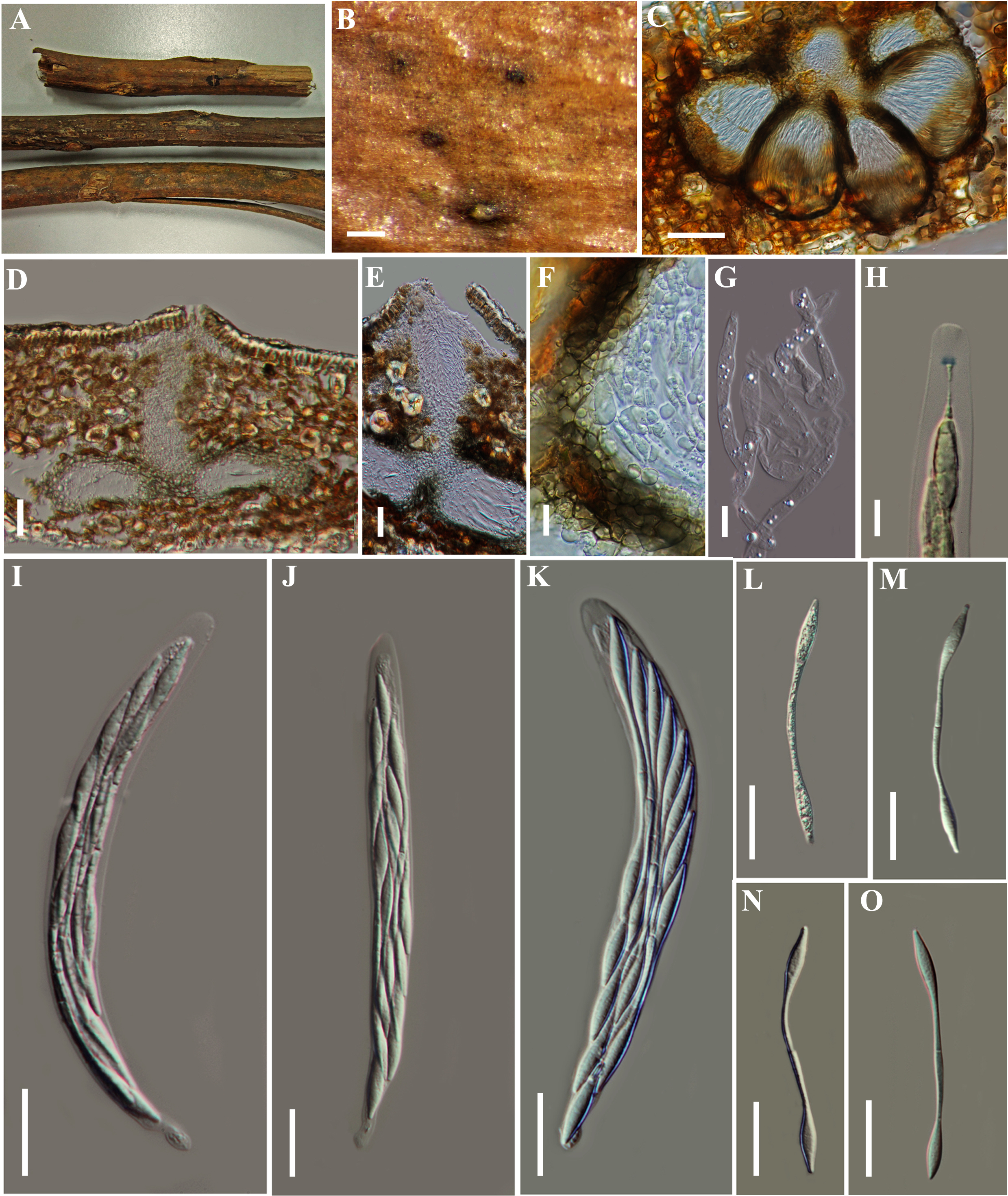
Fig. 1 Vialaea mangiferae (MFLU13–0342, holotype). A Dead twigs of Mangiferaindica. B Appearance of pseudostromata on the natural substrate. C Horizontal section of ascomata with central neck. D Vertical section of ascomata with central neck. E Periphyses. F Peridium. G Paraphyses. H Apical ring, blueing at the base in Melzer’s reagent. I–K Asci in water. L–O Ascospores. Note: The central ostiolar canal and septum of ascospore arrowed in 4 and 14. Scale bars: C = 200 µm, D–F = 50 µm, G–H = 10 µm, I–O = 20 µm.
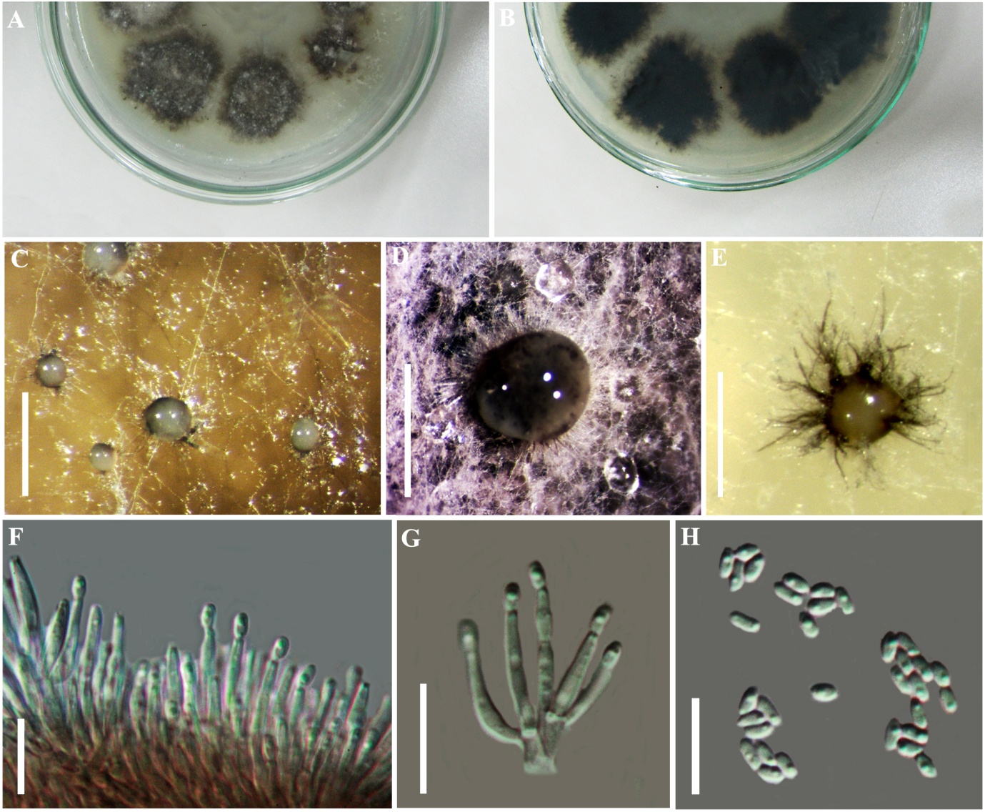 Fig. 2 Vialaea mangiferae (Coelomycetous asexual state, ex-type). A Culture from above. B Culture from below. C–E Conidiomata forming on WA. F Conidia forming on phialides attached to conidiophores. G Conidia attached to lageniform intercalary cells. H Conidia. Scale bars: C–F = 500 µm, D = 1 mm, F = 10 µm.