Vandijckomycella joseae Hern.-Restr., L. W. Hou, L. Cai & Crous, sp. nov.
MycoBank number: MB 833208; Index Fungorum number: IF 833208; Facesoffungi number: FoF 12628; Figure 11
Etymology. Named in honour of the first female President (2015–2018) of the Royal Dutch Academy of Arts and Sciences (KNAW), José F.T.M. van Dijck, who collected the soil sample from which the ex-type strain was isolated.
Typus. The Netherlands. North Holland province, Amsterdam, isolated from garden soil, Mar. 2017, J.F.T.M. van Dijk (holotype designated here CBS H-24112; living ex-type culture CBS 143011 = JW 1073).
Conidiomata pycnidial, produced on the agar surface, scattered or aggregated, solitary, (sub-)globose, confluent and irregularly-shaped with age, pale brown, covered in abundant long and thin mycelium hair, 150–340 × 130–250 µm; with 1–2 slightly papillate or non-papillate ostioles, sometimes elongated to a short neck; pycnidial wall pseudoparenchymatous, 3–5 layers, 13–25 µm thick, outer layers composed of brown, flattened, polygonal cells of 10–23 µm diam. Conidiogenous cells phialidic, hyaline, smooth, globose, ampulliform, lageniform or subglobose, 5–8(–9.5) × 4–8 µm. Conidia ellipsoidal to oblong, smooth- and thin-walled, hyaline, aseptate, 3.5–5.5 × 2–2.5 µm, (1–)2(–3)-guttulate. Conidial matrix whitish.
Culture characteristics. Colonies after 7 d at 25 °C, on OA reaching 75–80 mm diam after 7 d, covered by woolly aerial mycelium, concentric circles, pale olivaceous grey, pink, pale greenish grey, whitish near the edge, margin regular; reverse concentric circles dark brown, pale brown, orange, and pale olivaceous. On MEA reaching 75–80 mm diam, aerial mycelium woolly, margin regular, pale olivaceous grey; reverse dark brown, reddish towards the periphery. On PDA reaching 75–80 mm diam, mar- gin regular, covered by felty aerial mycelium, pale olivaceous grey or olivaceous grey, with whitish parts near the centre or through the plate; reverse zonate, orange to red-dish, brown and yellow. NaOH spot test: a coral discolouration on OA.
Additional specimen examined. The Netherlands. North Holland province, Amsterdam, isolated from garden soil, Mar. 2017, J.F.T.M. van Dijk, CBS 144948 = JW 1068.
Notes. The new genus Vandijckomycella is introduced to accommodate two new species isolated from soil samples which form an independent lineage in Didymellaceae, being clearly separated from other genera (Figure 1). Based on the phylogenetic analysis, V. joseae forms a distinct lineage which is distant from the nearest species V. snoekiae, and chiefly differs on tub2 and rpb2 sequences. Morphological differences between V. joseae and V. snoekiae are discussed under the latter species. Vandijckomycella joseae is characterised by producing pycnidia with longer whitish hyphal outgrowths, and with elongated necks.
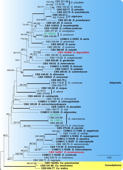
Figure 1. Phylogenetic tree generated from the maximum-likelihood analysis based on the combined ITS, LSU, tub2 and rpb2 sequence alignment of Didymellaceae members. The RAxML bootstrap support values (BS), Bayesian posterior probabilities (PP), and parsimony bootstrap support values (PBS) are given at the nodes (BS/PP/PBS). BS and PBS values represent parsimony bootstrap support values >50 %. Full supported branches are indicated in bold. The scale bar represents the expected number of changes per site. Ex-type strains are represented in bold. Strains obtained in the current study are printed in green; among them, whilst strains that represent new taxa are printed in red. Some of the basal branches were shortened to facilitate layout (the fraction in round parentheses refers to the presented length compared to the actual length of the branch). The tree was rooted to Coniothyrium palmarum CBS 400.71 and Leptosphaeria doliolum CBS 505.75.
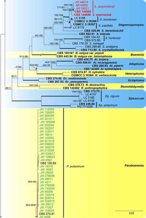
Figure 1. Continued.
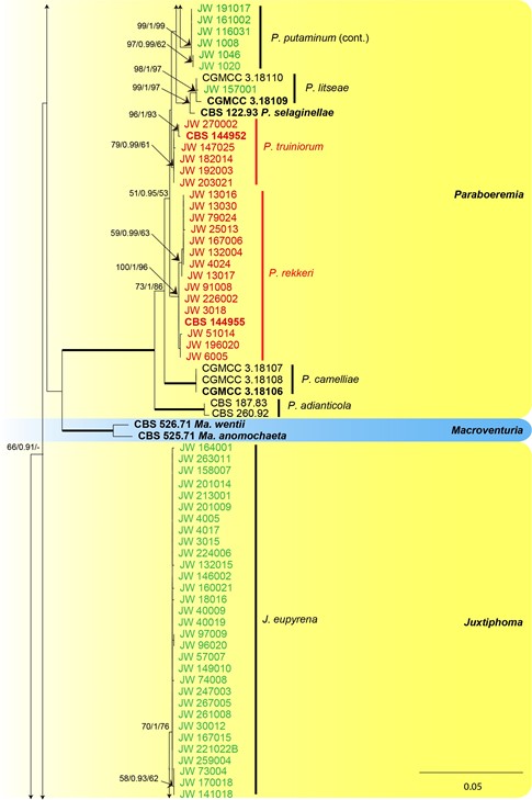
Figure 1. Continued.
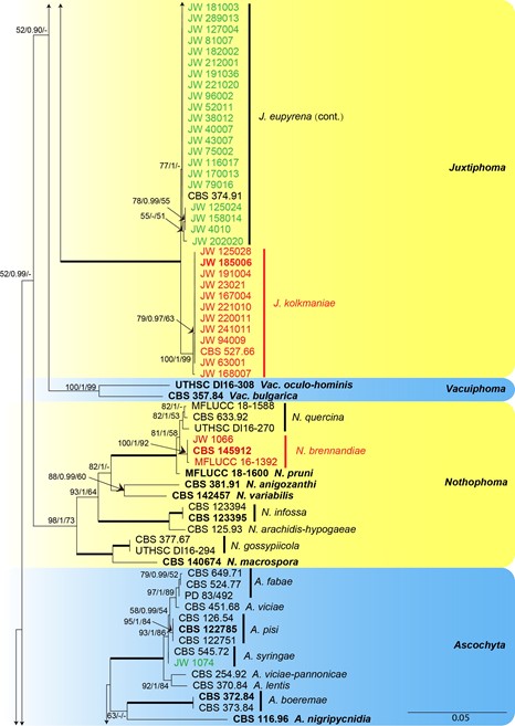
Figure 1. Continued.
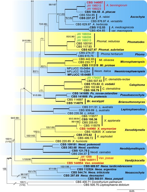
Figure 1. Continued.
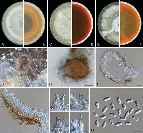
Figure 11. Vandijckomycella joseae (CBS 143011). A, B Colony on OA (front and reverse) C, D colony on MEA (front and reverse) E, F colony on PDA (front and reverse) G, H pycnidia forming on OA I, J section of pycnidial wall K–N conidiogenous cells O conidia. Scale bars: 100 µm (H); 20 µm (I); 10 µm (J); 5 µm (K–O).
