Trochilispora schefflerae VP Abreu, AWC Rosado & OL Pereira, in Hyde et al., Fungal Diversity: 10.1007/s13225-019-00429-2, [171] (2019)
MycoBank number: MB 555485; Index Fungorum number: IF 555485; Facesoffungi number: FoF 04860
Etymology – Name derived from its host genus, Schefflera.
Holotype – VIC 44384
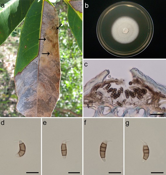
Asexual morph Conidiomata acervular, epiphyllous, scattered and occasionally confluent, subepidermal in origin, erumpent, rounded to oval in outline, 49–88 × 79–235 μm diam., unilocular, brown or black, basal stroma thick, of textura angularis, cells thick-walled and almost colourless; lateral walls 3–5 cells thick, cells thick-walled, pale brown to brown. Conidiophores cylindrical to subcylindrical (8.5–15.5 × 1.5–2 μm), formed in the concavity of the conidioma, unbranched, hyaline, smooth-walled. Conidiogenous cells discrete, annellidic with 2 annellations (3.5–11.5 × 1.5–3 μm), hyaline, thin- and smooth-walled. Conidia fusiform, straight or slightly curved, concolourous, smooth, bearing apical appendage, and basal appendage absent; 3-septate (13–19 × 3.5–5 μm), bearing: [basal cell obconic to conic, hyaline to subhyaline, smooth and thin-walled, 2–4 μm long; two median cells doliiform, 8.5–12.5 μm long, smooth, concolourous, brown, septa darker than the rest of the cell (second cell from base brown, 3.5–5.5 μm long; third cell brown, 4.5–7 μm long); apical cell 2–3.5 μm long, hyaline to subhyaline, subconical to hemispherical, thin- and smooth- walled; with 1 tubular apical appendage, arising from the apical crest, not centric, unbranched, filiform, 2–6.5 μm long; basal appendage absent], or 4-septate (15.5–21 × 4–5 μm), bearing: [basal cell obconic to conic, hyaline to subhyaline, smooth and thin-walled, 2–5 μm long; three median cells doliiform, 10–13 μm long, smooth, concolourous, brown, septa darker than the rest of the cell (second cell from base brown, 4–6 μm long; third cell brown, 2.5–4 μm long; fourth cell brown, 2.5–4.5 μm long); apical cell 2.5–3.5 μm long, hyaline to subhyaline, subconical to hemisphaerical, thin- and smooth-walled; with 1 tubular apical appendage, arising from the apical crest, not centric, unbranched, filiform, 2.5–7.5 μm long; basal appendage absent. Sexual morph Undetermined.
Culture characteristics – Colonies cultured on PDA reaching 38 mm diam. after 1 wk at 25 °C with a photoperiod of 12 h; regularly margins; with dense aerial mycelium; white; colonies fertile (Fig. 124b). Colonies cultured on MEA reaching 40 mm diam. after 1 wk at 25 °C with a photoperiod of 12 h; regularly and submerged margins; with scarce and sebaceous aerial mycelium; pale yellowish; colonies fertile.
Material examined – Brazil, Minas Gerais, Paraopeba, Floresta Nacional de Paraopeba (FLONA-Paraopeba), on leaves of Schefflera morototoni (Araliaceae), 30 January 2016, V.P. Abreu & O.L. Pereira (VIC 44384, holotype), ex-type living culture (COAD 2371).
GenBank numbers – ITS: MH128360, LSU: MH084761, TEF1-a: MH231216, TUB2: MH231215.
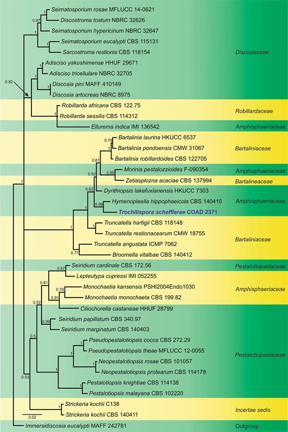
Notes – Trochilispora is introduced as a new genus based on morphology and phylogenetic support (LSU and ITS sequence data). Based on phylogenetic analyses, Trochilispora schefflerae COAD 2371 grouped in a well-supported clade, including Hymenopleella hippophaeicola CBS 140410 (Fig. 125), but different genera can be grouped in the same clade as, for example, Morinia and Zetiasplozna; Truncatella and Broomella; among others. Unfortunately, Jaklitsch et al. (2016) did not observe the asexual morph of Hymenopleella hippophaeicola, but the authors cite the Hymenopleella sollmannii species reported by Shoemaker and Müller (1965). The phylogenetic position of the Trochilispora family is still unclear. Trochilispora schefflerae COAD 2371 differs from Hymenopleella sollmannii by having conidia formed in conidiomata acervular with lateral walls 3–5 cells thick of brown hyphae; conidiophores smaller; conidiogenous cells discrete, annellidic with 2 annellations; conidia fusiform, straight or slightly curved, 3–4-septate, with medium brown central cells and hyaline to subhyaline end cells, apical cell with an appendage tubular, filiform, single, not centric, unbranched, not septum and basal cell without appendage basal. Our phylogenetic tree was built using LSU and ITS data, and morphological features corroborated that our isolate represents a new genus and a new species belonging to Amphisphaeriaceae.

Fig. 1 Trochilispora schefflerae (VIC 44384, holotype). a Leaves of Schefflera morototoni in Floresta Nacional de Paraopeba, state of Minas Gerais, Brazil (the arrows indicate the reproductive structures of the fungus). b Colony on PDA after 1 wk at 25 °C with a photoperiod of 12 h in the dark in Petri dishes (90 9 15 mm) (COAD 2371). c Cross section of the conidioma. d–g Conidia. Scale bars: c = 50 μm, d–g = 10 μm.

Fig. 2 Phylogram generated from Bayesian Inference analysis based on combined ITS and LSU sequence data for several closely related genera in Amphisphaeriales. Sequence data of ex-type or ex- epitype cultures are taken from Senanayake et al. (2015), and the closest hits of the GenBank database were included in this study. The combined genes sequence analysis included 40 taxa, which comprise 1405 characters (591 characters for ITS, 814 characters for LSU), and outgroup taxon Immersidiscosia eucalypti (MAFF 242781). Bayesian posterior probability is indicated at the nodes, and values equal to or greater than 0.95 are in bold. Isolate numbers are indicated after species names. The ex-type or ex-epitype strains are in bold and black. The newly generated sequence is indicated in bold and blue
