Trechispora taiwanensis S.L. Liu, S.H. He & L.W. Zhou.
MycoBank number: MB 559902; Index Fungorum number: IF 559902; Facesoffungi number: FoF 12882;
Description
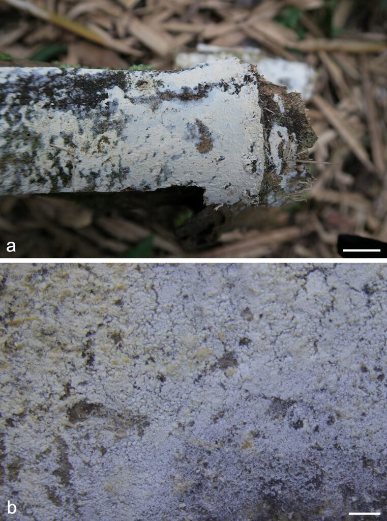
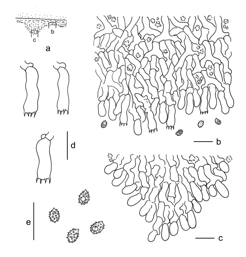
Basidiomes annual, resupinate, effused, thin, soft, fragile, loosely attached to the substrates, up to 10 cm long, 5 cm wide. Hymenophore smooth to grandinioid with numerous small aculei, farinaceous, cream when fresh, cream to straw-yellow with age, finely cracked with age. Margin thinning out as byssoid, 2–3 mm wide. Hyphal system monomitic; generative hyphae with clamp connections. Subicular hyphae long-celled, hyaline, thin-walled, frequently branched and septate, subparallel, 2.5–4.5 µm in diam. Aculei composed of a central core of compact hyphae and subhymenial and hymenial layers; generative hyphae distinct, hyaline, thin or thick-walled, moderately branched, smooth, subparallel, 2.5–5.5 μm in diam. Cystidia absent. Crystals present only in subiculum, bipyramidic, aggregated. Basidia cylindrical with a slight median constriction, hyaline, thin-walled, with four sterigmata and a basal clamp connection, 13–16 × 4–5.5 µm; basidioles in shape similar to basidia, but slightly smaller. Basidiospores ellipsoid, hyaline, thin-walled, aculeate, inamyloid, indextrinoid, acyanophilous, sometimes with one oil drop in the protoplasm, 3–4(–4.2) × 2–2.8(–3.2) µm, L = 3.3 µm, W = 2.5 µm, Q = 1.2–1.5 (n = 60/2).
Material examined: CHINA, Taiwan, Nantou County, Lianhuachi Nature Reserve, on dead bamboo, 6 Dec. 2016, S.H. He, He 4571 (holotype in BJFC 024012).
Distribution: CHINA
Notes: Macromorphologically, T. taiwanensis resembles T. laevis, but T. laevis differs in straight or concave basidiospores at the ventral side (Larsson 1996). In addition, T. taiwanensis is so far only known on bamboo in subtropical Asia, whereas T. laevis grows on coniferous wood in North Europe (Larsson 1996).

Fig. 1. Basidiomes of Trechispora taiwanensis (He 4571, holotype). — Scale bars: a = 1 cm; b = 1 mm.

Fig. 2. Microscopic structures of Trechispora taiwanensis (drawn from the holotype). a. Vertical section through basidiomes showing position of b and c; b. Section through base of a spine; c. Section through apex of a spine; d. Basidia; e. Basidiospores. — Scale bar = 10 μm.
