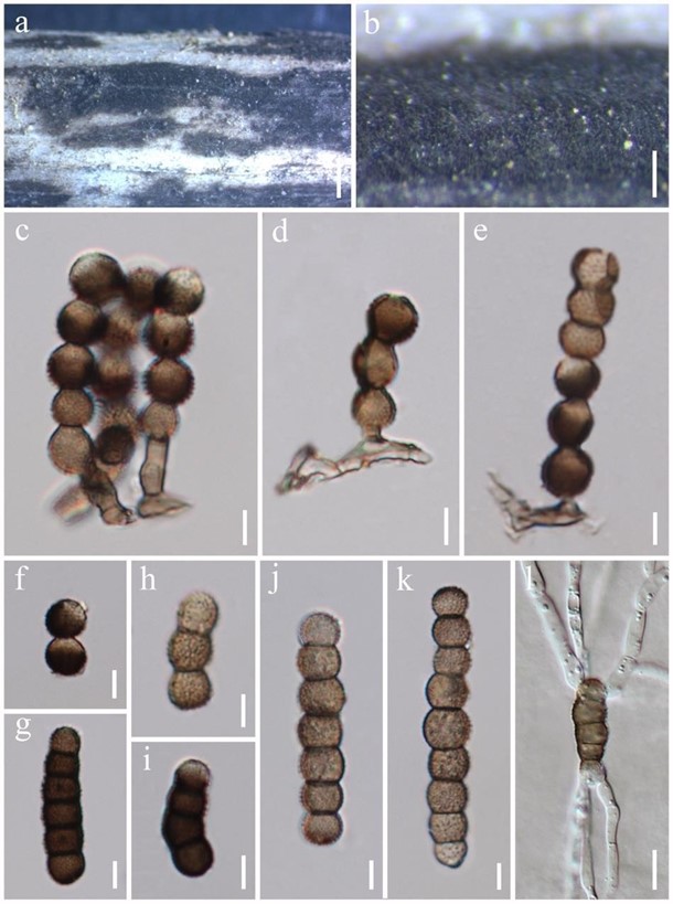
Torula thailandica N.G. Liu, Jian K. Liu & K.D. Hyde, sp. nov. Fig. 158
MycoBank number: MB 557091; Index Fungorum number: IF 557091; Facesoffungi number: FoF 06655.
Etymology – Referring to the country in which the species was collected. Holotype – MFLU 19-2856.
Saprobic on decaying wood. Sexual morph: Undetermined. Asexual morph: Colonies effuse on host, powdery, black. Conidiophores 1.4–4.2 μm wide, micronematous to semi-macronematous, mononematous, solitary, erect, subhyaline to paler brown smooth to minutely smooth, thin-walled, consisting of 1–2 cells, without apical branches, with ampulliform cells, arising from prostrate hypha, sometimes absent. Conidiogenous cells 2–9.3 × 2–3.9 μm (x̅ = 5.3 × 3 μm, n = 6), monoblastic, terminal, dark brown to black, smooth to minutely verruculose, thick-walled, ellipsoid to coronal. Conidia 14–23(–52.5) × 4.5–6.6 μm (x̅ = 18.1 × 5.6 μm, n = 30), solitary, acrogenous, phragmosporous, rarely in branched chains, chiefly subcylindrical, greyish-brown to blackish- brown, 2–8-septate, composed of moniliform cells, slightly constricted at some septa, minutely verruculose to verruculose. Conidial secession schizolytic.
Culture characteristics – Conidia germinating on water agar within 24 h. Germ tubes produced from both sides. Colonies on MEA velvety, circular, greyish brown in the center, greyish white in the periphery from above.
Material examined – Thailand, Phrae Province, Rong Kwang, on decaying wood, 10 January 2018, N.G. Liu, N002, (MFLU 19-2856, holotype); ex-type living culture, GZCC 20-0011.
GenBank numbers – ITS: MN907426; LSU: MN907428; SSU: MN907427.
Notes – Torula thailandicum is similar to T. chiangmaiensis and T. pluriseptata. However, conidia of T. thailandicum have less septa (2–8-septate vs 4–12-septate and 3–10-septate) and are shorter (14–23 μm vs. 25.5–70 μm and 23.5–36 μm) than those of T. chiangmaiensis and T. pluriseptata. Phylogenetic analyses also confirmed they are distinct species.

Figure 158 – Torula thailandica (MFLU 19-2856, holotype). a, b Colonies on the host. b Mass of conidia. c–e Conidiophores bearing conidia. f Conidiogenous cells. g–k Conidia. l Germinated conidia. Scale bars: a = 1000 μm, b = 200 μm, c–k = 5 μm, l = 10 μm.
