Tirisporella E.B.G. Jones, K.D. Hyde & Alias, Can. J. Bot. 74(9): 1489 (1996)
MycoBank number: MB 27659; Index Fungorum number: IF 27659; Facesoffungi number: FoF 02139; 1 species with sequence data.
Type species – Tirisporella beccariana (Ces.) E.B.G. Jones, K.D. Hyde & Alias
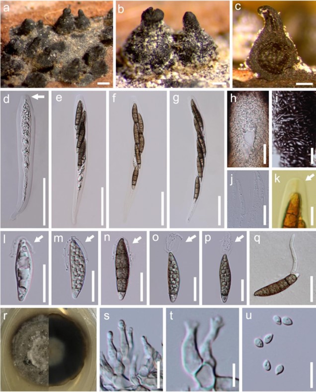
Notes – The monotypic genus Tirisporella was introduced by Jones et al. (1996) to accommodate the type species T. beccariana (≡ Sphaeria beccariana), which was frequently encountered on intertidal petioles of Nypa fruticans (mangrove palm). This genus has historically been classified in the Loculoascomycetes incertae sedis, and Pleosporales incertae sedis because of the bitunicate-like asci (Jones et al. 2009), while the familial placement was confirmed and assigned into Sordariomycetes based on phylogenetic analysis (Suetrong et al. 2015). The most obvious characters of Tirisporella are the first basal septum delimiting a hyaline to light-coloured basal cell, the remaining cells brown and verrucose ascospores with apical appendages. During examination of intertidal fungi from Nypa fruticans, fresh collections of T. beccariana were made and enabled a re-description and illustration of the fungus, and herein we provide an updated phylogenetic tree with all strains of this order. Tirisporella beccariana is illustrated in this entry.

Figure 246 – Tirisporella beccariana (Material examined – THAILAND, Ranong, Ngao (Ranong) Mangrove Forest Research Center, on intertidal petiole of Nypa fruticans Wurmb., 7 December, 2016, S.N. Zhang, SNT82, living culture MFLUCC 18-1572, specimen voucher MFLU 18-1582, HKAS 97482; THAILAND, Prachuap Khiri Khan, Pak Nam Pran, on intertidal petiole of Nypa fruticans, 2 December, 2016, S.N. Zhang, SNT102, living culture MFLUCC 18-1571, specimen voucher MFLU 18-1584; THAILAND, Krabi, Pali, on intertidal petiole of Nypa fruticans, 30 August, 2017, S.N. Zhang, SNT203, specimen voucher MFLU 18-1585, HKAS 97483). a, b Appearance of ascomata on host surface with ostioles. c Vertical section through the ascoma. d-g Asci. h Ostiole with periphyses. i Structure of peridium. j Paraphyses. k Apex of ascus in Lugol’s iodine, with a J-, apical ring. l-p Ascospores. q Germinating spore. r Colony on PDA. s-u Asexual morph structure in culture. Scale bars: a = 500 μm, b, c = 200 μm, d-g = 50 μm, h-j, l-q = 20 μm, k, s, u = 10 μm, t = 5 μm.
Species
