Synaptospora petrakii Cain, Beih. Sydowia 1: 5 (1957) [1956]
MycoBank number: MB 306588; Index Fungorum number: IF 306588; Facesoffungi number: FoF 10048; Fig. 64
Saprobic on decorticated wood. Sexual morph: Ascomata 200–350 × 250–450 µm (x̄= 300 × 350 µm, n = 5), perithecial, superficial, gregarious to scattered, globose to subglobose, carbonaceous, with ostiole, black, verrucose, surrounded by sparse, brown, septate setae, 3.5–7 µm wide, with a rounded apex. Subiculum ascomata seated on sparse, brown, septate, branched, friable hyphae, 5.5–7.5 µm wide. Peridium 30–75 µm (x̄=55 µm, n=20) wide, comprising two layers, outer layer carbonaceous, composed of brown cells of textura angularis; inner layer membranaceous, composed of hyaline cells of textura prismatica. Paraphyses 2.5–8.5 µm wide, septate, slender to cylindrical, evanescent. Asci (70–)80–95(–100) × 7.5–10 µm (x̄=85 ×8 µm, n=30), normally 2-spored, unitunicate, cylindrical, broadly rounded and thickened at the apex, long pedicellate. Ascospores (20–)22–24(–26) × 4–8 µm (x̄= 23 × 6 µm, n = 50) for four cells, uni-seriate, subglobose to ellipsoidal, hyaline and aseptate when young, gradually becoming cylindrical to oblong, dark brown, usually 2–4-septate, distinctly constricted at the septa, the cells swollen, ends broadly rounded, straight to slightly curved, smooth-walled, with a large guttule in each cell, breaking into part spores. Part spores 6–9 × 4.5–7 µm (x̄= 7.5 × 5.5 µm, n = 50), hyaline to dark brown, globose to ellipsoidal, aseptate, smooth-walled, with a large guttule, discharging through constricting septa. Asexual morph: Undetermined (adapted from Cain 1957b).
Material examined – Canada, Ontario, north of Mississauga, in willow grove by Credit River on Creditview Road, on decaying logs, 11 November 1987, L.A. Novak (TRTC- 51203); Canada, Ontario, Muskoka district, Haliburton, 11 km south of Dorset, on decorticated wood, October 1975, D. Tighe (TRTC-51205).
Known hosts and distribution – On dead decorticated log of Betula papyrifera in Canada (type locality) (Cain 1957b).
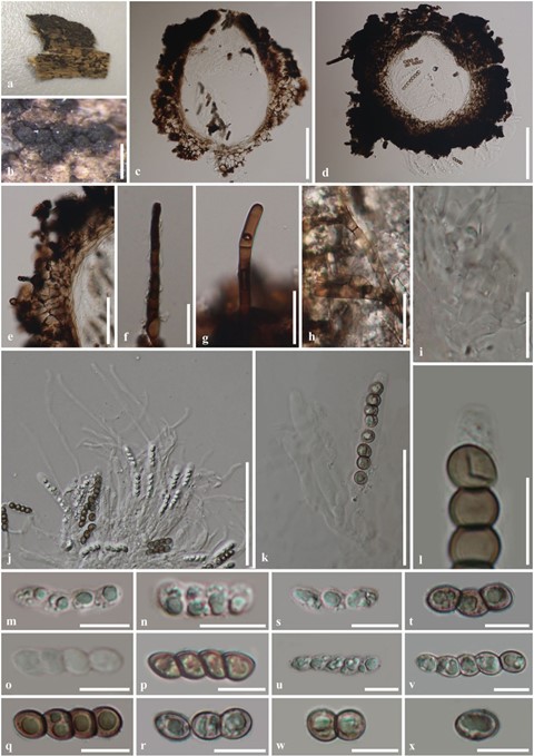
Notes – The holotype material (TRTC-32168) was collected by Cain (1957b) in Canada. We were unable to obtain the type material and, therefore, we re-examined two authentic samples, TRTC-51203 and TRTC-51205, collected in Canada and determined by M. Matzer (mentioned in the label of material). Ascospores of Synaptospora were described as ellipsoidal to globose and fused in groups when mature (Cain 1957b; Barr 1990; Huhndorf et al. 1999b). We could not find mature ascospores fused in groups in TRTC-51203 and TRTC-51205. However, we found that the ascospores were initially hyaline, ellipsoidal to oblong or cylindrical and gradually formed distinct septa, and finally, the septa split the brown ascospores into individual globose to subglobose part spores (Fig. 64m–r). Fresh collections of Synaptospora species are required to determine the nature of ascospores and the placement of the genus.

Fig. 64 Synaptospora petrakii: a–c, e, f, h–o, q–x (TRTC- 51203); d, g, p (TRTC-51205). a Herbarium material. b Ascomata on host. c, d Ascoma in cross section. e Peridium. f, g Setae. h Hyphae of subiculum. i Septate paraphyses. j Asci with paraphyses. k Asci. l Apex of ascus (stained in Melzer’s reagent). m–q, s–v Immature to mature ascospores (m–q 3-septate; s–t 2-septate; u–v 4-septate). r, w Ascospores breaking into individual cells. x Individual spore cells. Scale bars: b = 500 µm, c. d. j = 100 µm, e. k = 50 µm, f–i = 20 µm, l–x = 10 µm
