Skeletocutis subchrysella B.K. Cui & Shun Liu
MycoBank number: MB 559466; Index Fungorum number: IF 559466; Facesoffungi number: FoF 10675;
Description
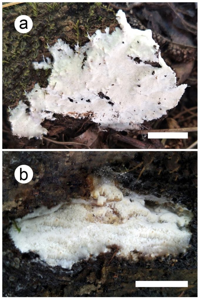
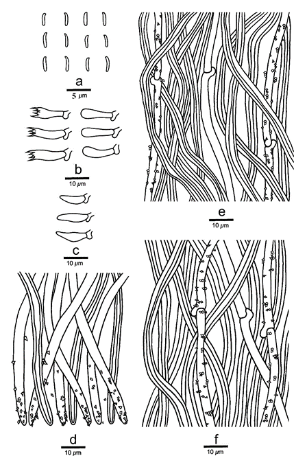
Fruiting body basidiocarps annual, resupinate, not easily separated from substrate, soft leathery, without odour or taste when fresh, becoming corky upon drying, up to 6 cm long, 1.5 cm wide, and 3 mm thick at center. Pore surface white to cream when fresh, becoming cream to cinnamon-buff upon drying; pores angular, 6–8 per mm; dissepiments slightly thick, entire to lacerate. Subiculum cream, corky, up to 0.5 mm thick. Tubes darker than poroid surface, corky, up to 2 mm long. Hyphal Structure hyphal system dimitic; generative hyphae with clamp connections, hyaline, thin-walled, dominant at dissepiment edge; skeletal hyphae with a wide to narrow lumen; IKI–, CB–, unchanged in KOH. Subiculum generative hyphae frequent, hyaline, thin-walled, unbranched and bearing fine crystals, 1.5–2.8 μm in diameter; skeletal hyphae dominant, thick-walled with a narrow lumen, flexuous, unbranched, interwoven, 2–4 μm in diameter. Tubes generative hyphae frequent, thin-walled, unbranched, usually covered by fine crystals, sharply pointed encrustations, especially at dissepiment edge, 1.5–2.5 μm in diameter; skeletal hyphae dominant, thick-walled with a wide to narrow lumen, unbranched, subparallel along the tubes, not agglutinated, 2–3.7 μm in diameter. Dissepiment edge dimitic with smooth skeletal hyphae, and dominant winding, encrusted generative hyphae. Cystidia absent, cystidioles abundant, bottle-shaped, with a conical apex, 8–13.8 × 2–3.6 μm. Basidia clavate, with a basal clamp connection and four sterigmata, 10.3–15.6 × 3.2–4.5 μm; basidioles in shape similar to basidia, but slightly smaller. Spores basidiospores allantoid, hyaline, thin-walled, smooth, IKI–, CB–, (2.6–)2.7–3.2 × (0.5–)0.7–1 μm, L = 2.93 μm, W = 0.8 μm, Q = 3.33–4.5(n = 60/2).
Material examined: CHINA, Sichuan Province, Shimian County, Liziping National Nature Reserve, on fallen angiosperm trunk, 14 September 2019, Cui 17748 (holotype, BJFC); Yunnan Province, Baoshan, Gaoligongshan Nature Reserve, on fallen angiosperm trunk, 8 November 2019, Cui 18141 (paratype, BJFC).
Distribution: CHINA. Sichuan Province, Shimian County, Liziping National Nature Reserve; Yunnan Province, Baoshan, Gaoligongshan Nature Reserve
Sequence data: Cui 17748 (ITS: MZ327278; nLSU: MZ348534), Cui 18141 (ITS: MZ327282; nLSU: MZ348538)
Notes: Skeletocutis subchrysella is characterized by its white to cream pore surface when fresh, cream to cinnamon-buff upon drying, and allantoid basidiospores (2.7–3.2 × 0.7–1 μm). In the phylogenetic tree, Skeletocutis subchrysella grouped with S. chrysella, S. delicata Niemelä & Miettinen, S. exilis Miettinen & Niemelä and S. kuehneri A. David. Morphologically, they all have allantoid basidiospores; but S. chrysella differs from S. subchrysella in its trimitic hyphal system and longer basidiospores (2.8–4.5 × 0.7–1 μm; Niemelä 1998); S. delicata and S. exilis differ by having larger pores (3–6 per mm in S. delicata, 3–5 per mm in S. exilis) and basidiopores (3.2–4.2 × 1.1–1.4 μm in S. delicata, 3.2–3.9 × 0.9–1.1 μm in S. exilis; Miettinen and Niemelä 2018); S. kuehneri differs by having thin and brittle basidiocarps and growth on dead wood of Picea and Pinus (David 1982). Skeletocutis bambusicola, S. lepida, S. mopanshanensis and S. yunnanensis were also discovered from Yunnan Province. Skeletocutis bambusicola differs from the new species by having smaller pores (8–11 per mm) and ellipsoid basidiospores (2.7–3.1 × 1.5–1.9 μm; Zhou and Qin 2012); S. lepida differs by having half-resupinate basidiocarps with ochraceous upper surface when dry (Korhonen et al. 2018); S. mopanshanensis differs by having larger pores (4–5 per mm) and basidiospores (4.7–6.6 × 3.2–4.5 μm; Wu et al. 2017); S. yunnanensis differs by having larger basidiospores (3.4–4.5 × 1–1.2 μm; Bian et al. 2016).

Fig. 1. a Skeletocutis cangshanensis (holotype) b S. subchrysella (holotype). Scale bars: a, b = 2 cm.

Fig. 2. Skeletocutis cangshanensis (holotype) a Basidiospores b Basidia and basidioles c Cystidioles d Section of dissepiment edge e Hyphae from trama f Hyphae from subiculum. Scale Bars: a = 5 µm, b-f = 10 µm.

Fig. 3. Skeletocutis subchrysella (holotype) a Basidiospores b Basidia and basidioles c Cystidioles d Section of dissepiment edge e Hyphae from trama f Hyphae from subiculum. Scale Bars: a = 5 µm; b–f = 10 µm.
