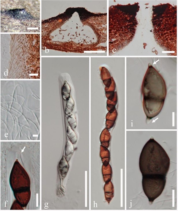
Salsuginea ramicola K.D. Hyde, Bot. Mar. 34(4): 316 (1991). Fig. 145
MycoBank number: MB 354934; Index Fungorum number: IF 354934; Facesoffungi number: FoF 08366.
Saprobic on decaying wood submerged in brackish waters in mangroves. Sexual morph: Ascomata 850–1100 × 320–400 μm (x̅ = 904 × 354 µm, n = 5), immersed, apical erumpent, solitary, subglobose to flask-shaped, smooth-walled, protruding papilla, conspicuous ostiole, dark to black. Ostiole central, cone-shaped, brown to black. Peridium 20–60 μm thick, comprising of light brown cells of textura porrecta, merging at the outside with the host, where textura angulata cells. Hamathecium comprising numerous, filiform, 2–3 μm wide, branched, septate, hyaline. Asci 300– 350 × 20–30 μm (x̅ = 379 × 27.3 µm, n = 20), 8-spored, fissitunicate, cylindrical-clavate, with an apical apparatus consisting of a large distinctive ocular chamber and prominent ring, sessile. Ascospores 60–75 × 20–30 μm (x̅ = 64 × 26 μm, n = 30), 1-seriate, obovoid, tapering toward sub- acute ends, brown, dark brown to black, 1-septate in lower third cell, constricted at the septa, colorless germ pore at each end, smooth walled. Asexual morph: Undetermined.
Material examined – Thailand, Ranong, 09º 58º N, 098º 37º E, in mangrove, on submerged decaying wood of Aegiceras cornicelarum in brackish water, October 1988, K.D. Hyde, BRIP 17102a, holotype).

Figure 145 – Salsuginea ramicola (BRIP 17102a, holotype). a Appearance of psuedoclypeus on host substrate. b Section of ascomata. c Ostiolatec. d Section of peridium. e Pseudoparaphyses. f Ocular chamber. g, h Asci with ascospores. i, j Ascospores with apical germ pores. Scale bars: a, b = 500 μm, c = 100 μm, d = 20 μm, e = 5 μm, f = 20 μm, g, h = 100 μm, i, j = 20 μm.
