Roussoella pseudohysterioides D.Q. Dai & K.D. Hyde, sp. nov. Index Fungorum number: IF552026
Etymology: Refers to the similarity with Roussoella hysterioides.
Holotype: MFLU 15–1209
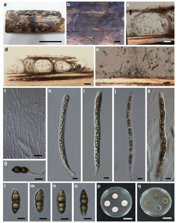
Saprobic on decaying bamboo culms. Sexual morph: Ascostromata forming under black area, up to 3–5 mm long and 1–2 mm wide, raised at maturity, ellipsoidal to irregular, black, coriaceous. Locules in vertical section 200–250 μm high, 170–350 μm diam., solitary to gregarious, subglobose to ellipsoidal, dark brown, with ostiolate opening. Peridium 15–25 μm wide, composed of dark brown to hyaline cells of compressed textura angularis, with upper wall 15–45 μm wide, darker, comprising host and fungal tissues. Hamathecium comprising dense, 2–3.5 μm wide, cellular pseudoparaphyses, indistinctly septate, anastomosing and branching at the apex, embedded in a gelatinous matrix. Asci 100–270 × 7–15 μm (x = 148.2 × 9.7 μm, n = 20),8-spored,bitunicate, cylindrical, with a short furcate pedicel, with an apical ocular chamber. Ascospores 12.5–20 × 4–6 μm (x = 16.1 × 5.2 μm, n = 20), uniseriate, fusiform-ellipsoidal, 1-septate, constricted at the septum, narrow at both ends, with longitudinally striate and verrucose wall ornamentation. Asexual morph: Undetermined.
Culture characters: Ascospores germinating on PDA within 24 h and germ tubes produced from lower end or both ends. Colonies circular, slow growing, 25 mm diam. in 30 days at 28–32 °C, under 12 h light/12 h dark, cottony, yellowish brown at the centre, light coloured and floccose at the periphery from above, yellow to dark brown to orange from below.
Material examined: THAILAND, Chiang Rai Province, Mae Fah Luang University, on dead culm of bamboo, 9 July 2013, Dong-Qin Dai DDQ00259 (MFLU 15–1209); Ibid. (KUN HKAS88717, isotype), ex-type living cultures, MFLUCC 13–0852, CBS 139992.
Notes: Roussoella pseudohysterioides is similar to R. hysterioides in having black stromatic ascomata, cylindrical asci and fusiform-ellipsoidal ascospores with longitudinal striations (Hyde et al. 1996). Roussoella pseudohysterioides, however, differs by its smaller ascospores (12.5–20× 4– 6 μm vs. 18–34 × 6–8 μm) (Hyde et al. 1996). Moreover, R. pseudohysterioides has verrucose ascospores, a character not observed in R. hysterioides. In the phylogenic tree (Fig. 19) R. pseudohysterioides is close to R. pustulata, while the branch length indicates that they are different species.
FIG. Roussoella pseudohysterioides (MFLU 15–1209, holotype). a, b Ascostromata developing on bamboo culm. c, d Vertical sections of ascostromata. e Peridium. f Branched pseudoparaphyses. g Germinating ascospore. h–k Asci containing eight ascospores. l–o Dark brown ascospores. p, q Cultures on MEA. Scale bars: a = 2 cm, b = 1 mm, c = 50 μm, d = 100 μm, e = 20 μm, f–k = 10 μm, l–o=5 μm, p, q = 25 mm