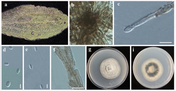
Remotididymella anthropophila anthropophila Valenz.-Lopez, Cano, Guarro & Stchigel, Stud. Mycol. 90: 35 (2017). Fig. 77
MycoBank number: MB 819991; Index Fungorum number: IF 819991; Facesoffungi number: FoF 06579.
Saprobic or pathogenic on leaves. Sexual morph: Undetermined. Asexual morph: Hyphae brown, smooth- and thin-walled, septate, Conidiomata pycnidial wall of textura angularis cells, 2– 5 layered, composed of subhyaline to pale brown flattened polygonal cells. Conidiogenous cells phialidic, hyaline, smooth-walled, ampulliform to globose, 5–6 μm diam. Conidia 3.7–6.0 × 1.7– 3.2 μm, cylindrical, hyaline, aseptate, smooth- and thin-walled, guttulate. Chlamydospores not observed.
Culture characteristics – Colonies reaching 35–40 mm diam on PDA after 5 days in the dark at 25 °C. Initially white and flattened with immersed mycelium with entire margin, with age become greyish brown. Reverse white and black in the middle with age become back.
Material examined – China, Yellow River Park, Shandong, on leaf spots belong to unknown host, 07 October 2017, Yuanyuan Hao – living culture, JZB380042.
Distributions – China, on unknown plant (this study). The USA, Texas, from human bronchial secretion.
GenBank numbers – ITS: MN648210, LSU: MN640405.
Notes – We isolated Remotididymella anthropophila from leaf spots. Morphology (Fig. 77) and phylogenetic analyses (Fig. 76) indicate that our collection is Remotididymella anthropophila. So far, this species is only reported on humans and this is the first report of R. anthropophila associated with a plant.

Figure 77 – Remotididymella anthropophila (JZB380042). a Material examined. b Appearance of polygonal cells associated with pycnidia. c Conidiogenus cell developing conidia d–f Conidia on PDA. g Mycelia h Upper view of the colony on PDA. i Reverse view of the colony on PDA. Scale bars: c, f = 10 µm, d, e = 3 μm.
