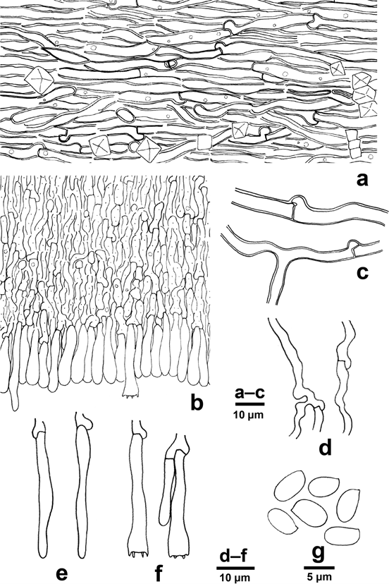
Quasiphlebia densa C.C. Chen & Sheng H. Wu, sp. nov. Figs. 24, 25
MycoBank number: MB 840762; Index Fungorum number: IF 840762; Facesoffungi number: FoF;
Typification: TAIWAN. Miaoli County, Taian Township, 19 km of East Branch of Dalu Forest Trail, 24°29ʹ N, 121°12ʹ E, on angiosperm branch, 23 Apr 2017, C.L. Wei, WEI 17-057 (holotype TNM F31834). GenBank: ITS = MZ637066; 28S = MZ637265; rpb1 = MZ748410; rpb2 = OK135986; tef1 = MZ913630.
Etymology: densa (Lat.), referring to the dense texture of the subiculum.
Description: Basidiocarps annual, effused, adnate, ceraceous, 0.4–1.2 mm thick in section. Hymenial surface pinkish-buff, clay-pink or yellowish-brown, not changing in KOH, smooth to tuberculate, not cracked; margin paler, fimbriate or arachnoid, thinning out. Hyphal system mono- mitic; generative hyphae nodose-septate; hyphal anastomosis occasionally present. Subiculum fairly uniform, with dense texture; hyphae horizontal, ± vertical near subhymenium, colorless, 3–9 μm diam, with 0.3–1.3 μm diam thick walls, moderately ramified, densely interwoven, agglutinated, ± flexuous. Cubic and pyramidal crystals frequently present in subiculum. Hymenial layer thickening, 50–150 μm thick, with compact texture, covered with yellow resinous materials, subhymenium clearly differentiated from subiculum, 30–130 μm thick; hyphae ± vertical, sub- colorless, 1.7–3.5 μm diam, distinctly narrower than those in subiculum, thin-walled, richly ramified, densely interwoven, agglutinated, flexuous, embedded in a gelatinized matrix. Cystidia 23–36 × 3–4 μm, cylindrical, infrequent, originating from hymenial layer, often projecting, thin-walled. Basidia 28–35 × 5–6 μm, narrowly clavate, thin-walled, 4-sterigmate, ± flexuous. Basidiospores ellipsoid, colorless, thin-walled, smooth, inamyloid, nondextrinoid, acyanophilous, mostly 4.2–5.4 × 2.3–3 μm. (4–)4.6–5.4(–5.8) × (2.3–)2.5–3(–3.5) μm, L = 5 μm, W = 2.8 μm, Q = 1.82 (n = 60) (holotype). (4.1–)4.2–4.9(–5.3) × (2.1–)2.3–2.7(–3) μm, L = 4.5 μm, W = 2.5 μm, Q = 1.82 (n = 30) (Wu 9304-33).
Other specimens examined: TAIWAN. Nanto County, Pilu, 24°14ʹ N, 121°17ʹ E, 2200 m, on angiosperm branch, 13 Apr 1993, S.H. Wu, Wu 9304–33 (TNM F14971).
UNITED STATES. GEORGIA: Clarke, Athens, University of Georgia Campus, on Quercus, 20 Feb 1988, H.H. Burdsall, Jr., HHB-12357 (CFMR).
Ecology and distribution: On angiosperm branch (e.g.,Quercus), Taiwan and S USA (Georgia), Feb, Apr.
Notes: The diagnostic features of Quasiphlebia densa are buff, thicker basidiocarps, smooth to tuberculate hyme- nial surface, dense subiculum with thick-walled, clamped hyphae, scattered cubic and pyramidal crystals, cylindrical leptocystidia, and ellipsoid basidiospores. Quasiphlebia densa resembles Antrodia griseoflavescens (Litsch.) Runnel, Spirin & K.H. Larsson, Flavophlebia sulfureoisabellina (Litsch.) K.H. Larss. & Hjortstamand and Phlebia tristis (Litsch. & S. Lundell) Parmasto by having smooth hymenial surface, clamped hyphae, and presence of cylindrical leptocystidia. However, Antrodia griseoflavescens has longer leptocystidia and cylindrical basidiospores (Runnel et al. 2019), while Flavophlebia sulfureoisabellina has subglobose basidiospores, and Phlebia tristis has allantoid basidiospores and larger leptocystidia (Bernicchia and Gorjón 2010).

Fig. 24 Basidiocarps of Quasiphlebia densa (WEI 17-057, holotype) in general and detailed views. Bars = 10 mm

Fig. 25 Micromorphological features of Quasiphlebia densa (drawn from WEI 17-057, holotype). a Part of vertical section from sub- iculum. b Part of vertical section from hymenial layer. c Subicu- lar hyphae. d Hyphae from hymenial layer. e Cylindrical cystidia. f Basidia. g Basidiospores
