Pteridiospora javanica Penz. & Sacc., Malpighia 11(9-10): 399 (1897)
= Apiospora carbonacea Rehm, Leafl. of Philipp. Bot. 8: 2945 (1916)
Index Fungorum Number: IF 200770; Facesoffungi number: FoF 000022
Saprobic on bamboo, visible as darkened areas on the host surface with several raised, cone-shaped, gregarious, easily broken fruiting bodies. Sexual state: Ascostromata 300–400 µm high, 500–700 µm diam., erumpent through host tissue, becoming superficial, conical, dark brown to black, solitary to gregarious, uniloculate, flattened at the base with ruptured reflexed tooth-like host remnants around the base, central ostiole, with small papilla. Peridium 30–70 µm wide, carbonaceous, composed of opaque dark cells, poorly developed at the base. Hamathecium comprising numerous 1–2 µm wide, narrow, trabeculate, filiform, branched, anastomosing, pseudoparaphyses, embedded in a gelatinous matrix. Asci (150–)160–190(–210) × (13–)16–18 μm ( = 181.5 × 16.5 µm, n = 25), 8-spored, bitunicate, fissitunicate, cylindric-clavate, short pedicellate, apically rounded with ocular chamber or surrounded by a faint ring, arising from the base of ascoma. Ascospores (31–)33–36(–39) × 8–9(–10) μm (= 34.2 × 8.3 μm, n= 30), overlapping, uni to bi-seriate, oblong to sub-fusoid narrowing towards apex, hyaline, 1-septate, constricted at the septum, upper cell longer than a lower cell, smooth-walled, guttulate, surrounded by a large appendage in the lower cell, with wide mucilaginous sheath. Asexual state: produced on sterilized bamboo pieces on WA or immersed in culture media. Conidiomata 130–260 µm high, 140–320 µm diam., black, globose on bamboo pieces, or forming conidiomata radiating outwards on cultures colony, erumpent to superficial, covered by vegetative hyphae, uniloculate, solitary to gregarious. Conidiomata wall 20–30 µm wide, composed of two layers of cells of equal thickness, outer layer of brown to dark brown textura intricata, inner layer of dark brown to black textura angularis. Conidiophores (2–)3–5(–8) × 1–2 µm, arising from the basal cavity around conidiomata, oblong to cylindrical or ampulliform, straight or slightly curved, hyaline, unbranched, aseptate. Conidiogenous cells integrated, phialidic. Conidia 2–3 × 2–3 µm ( = 2.4 × 2.1 µm, n = 45), globose to subglobose, forming chains, hyaline, aseptate, smooth-walled.
Culture characters – Colonies on PDA 51–53 mm diam. after 4 weeks at 25–30 ◦C, white at the edge, with fluffy radiating, grey to dark grey with small to large hyaline or black water drops and yellowish white fluffy in the centre; reverse white to pale yellowish at the edges, becoming grey to brownish grey or brownish orange in the middle with strongly radiating outwards colony, dense, circular to irregular shape, umbonate or raised with droplets in the centre, dull with entire edge, floccose, slightly radiated in the upper with strongly radiating in the lower, forming small black conidiomata radiating outwards after 8 weeks, non-pigmented.
Material examined – INDONESIA. Java: Tjibodas, on dead culms of Bambusae (Poaceae), 4 March 1896, no. 132 (PAD, holotype of Pteridiospora javanica); PHILIPPINES. Laguna, Los Baños, summit of Mt. Maquiling,on dead Schizostachyum
[Bambuseae] (Poaceae), June 1914, Baker_3427a (S_F7823, type of Apiospora carbonacea Rehm); THAILAND. Chiang Rai Province: Muang District, Khun Korn Waterfall, on dead stem of bamboo (Poaceae), 5 September 2010, R. Phookamsak RP0075, (MFLU 11-0195, epitype of Pteridiospora javanica designated here, isoepitype in PDD); ex epitype living culture = MFLUCC 11-0159 = ICMP; ibid., 17 December 2010, R. Phookamsak (MFLU 11-0231); living culture = MFLUCC 11-0195 = ICMP.
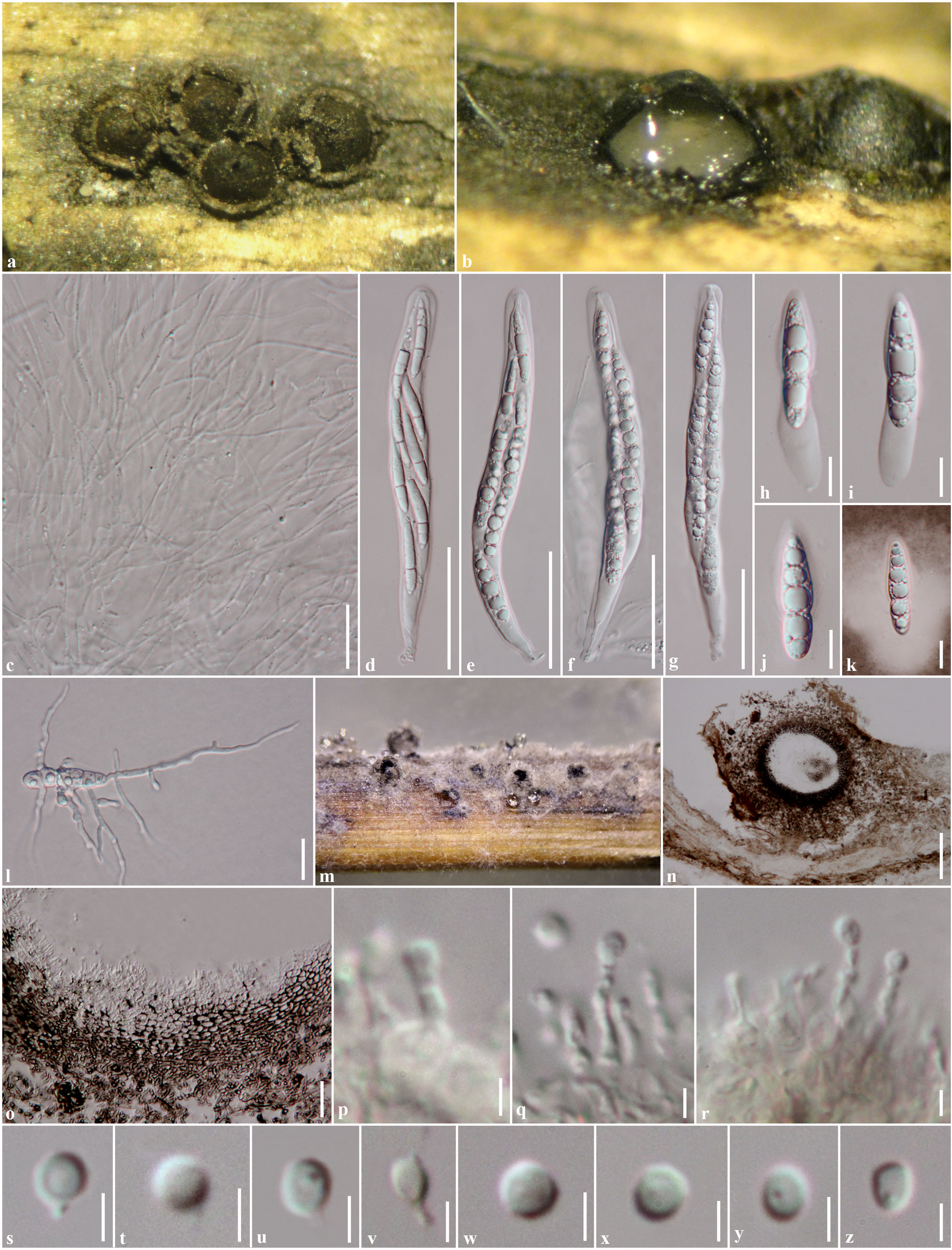
Figures of Pteridiospora javanica (MFLUCC11-0159) a. Ascostromata on host surface. b. Section through ascostroma. c. Pseudoparaphyses. d–g. Asci. h–j. Ascospores. k. Ascospore stained in Indian ink to show sheath. l. Germinating ascospore. m. Conidiomata forming on bamboo pieces on WA after 8 weeks. n. Section through conidioma. o. Section through conidioma wall. p–r. Conidiophores and conidiogenous cells. s–z. Conidia. Scale bars: n = 100 μm, p = 100 μm, d–g = 50 μm, c, l, o = 20 μm, h–k = 10 μm, p–z = 2 μm.
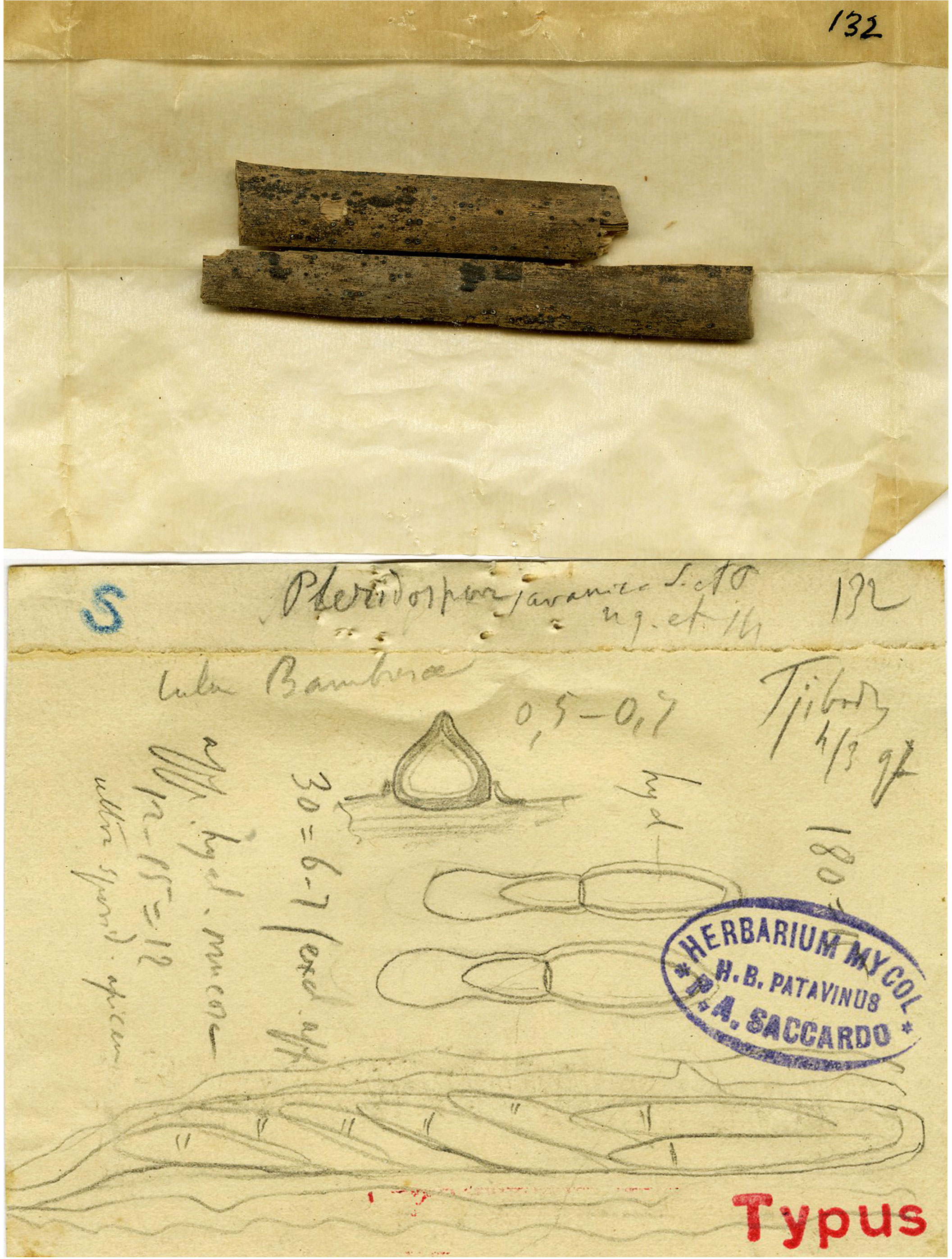
Figures of Iconotype of Pteridiospora javanica from PAD herbarium
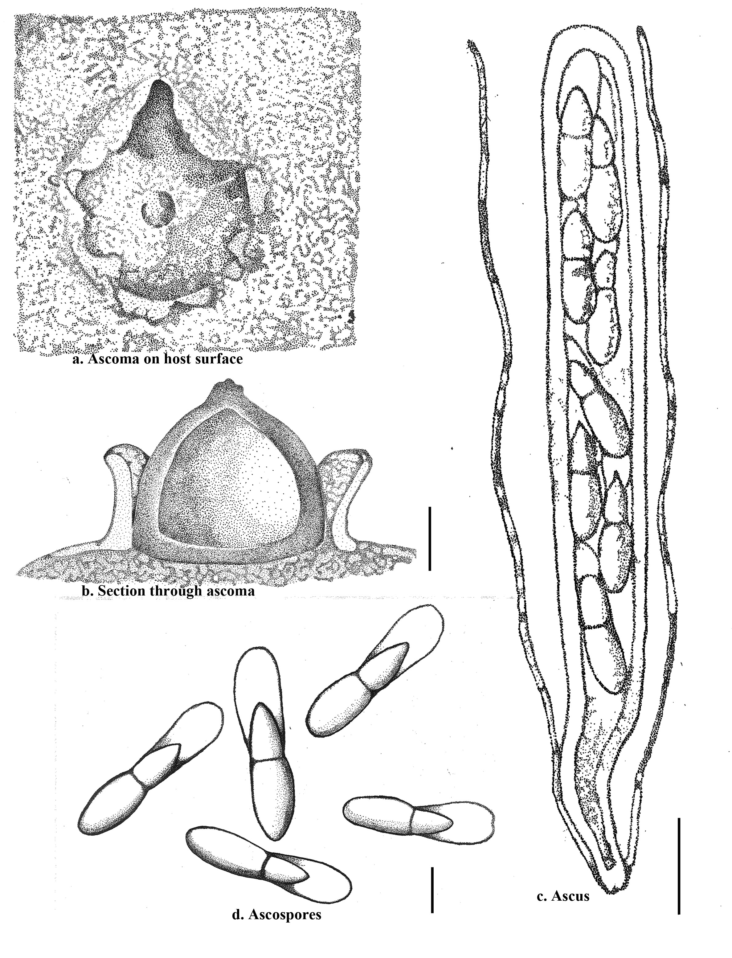
Figures of Pteridiospora javanica, redrawn from Penzig & Saccardo (1904). a. Ascostroma on host surface. b. Section through conical ascostroma. c. Ascus and pseudoparaphyses. d. Ascospores. Scale bars: a = 200 μm, b = 20 μm, c = 10 μm.